AI Approach Lowers Radiation Exposure from CT Imaging
|
By MedImaging International staff writers Posted on 06 Jul 2019 |

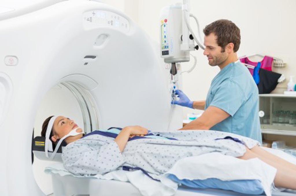
Image: Research shows machine learning has the potential to advance medical imaging, particularly CT scanning, by reducing radiation exposure and improving image quality (Photo courtesy of Axis Imaging News).
Engineers at the Rensselaer Polytechnic Institute (Troy, NY, USA) worked along with radiologists at Massachusetts General Hospital (Boston, MA, USA) and Harvard Medical School (Boston, MA, USA) to demonstrate that machine learning has the potential to vastly advance medical imaging, particularly computerized tomography (CT) scanning, by reducing radiation exposure and improving image quality. The team believes that their new research findings make a strong case for harnessing the power of artificial intelligence (AI) to improve low-dose CT scans.
Over the past several years, there has been significant focus on low-dose CT imaging techniques to alleviate concerns over patient exposure to X-ray radiation associated with widely used CT scans. However, reducing radiation can affect image quality. Engineers across the world have attempted to solve this problem by designing iterative reconstruction techniques to help sift through and remove interferences from CT images. However, the drawback is that these algorithms sometimes remove useful information or falsely alter the image.
In the latest research, the team attempted to address this persistent challenge by using a machine-learning framework. The developed a dedicated deep neural network and compared their best results to the best of what three major commercial CT scanners could produce with iterative reconstruction techniques. The researchers were looking to determine how the performance of their deep learning approach compared to the selected representative iterative algorithms currently being used clinically. They found that the deep learning algorithms developed by the Rensselaer team performed as well as, or better than, those current iterative techniques in an overwhelming majority of cases.
The researchers also found that their deep learning method was much quicker and allowed the radiologists to fine-tune the images according to clinical requirements. The positive results were realized without access to the original, or raw, data from all the CT scanners, and a more specialized deep learning algorithm is likely to perform even better if original CT data is made available, according to the researchers. They believe that these results confirm that deep learning could help produce safer, more accurate CT images while also running more rapidly than iterative algorithms.
“Radiation dose has been a significant issue for patients undergoing CT scans. Our machine learning technique is superior, or, at the very least, comparable, to the iterative techniques used in this study for enabling low-radiation dose CT,” said Ge Wang, the Clark & Crossan Endowed Chair Professor of biomedical engineering at Rensselaer, and a corresponding author on this paper. “It’s a high-level conclusion that carries a powerful message. It’s time for machine learning to rapidly take off and, hopefully, take over.”
Related Links:
Rensselaer Polytechnic Institute
Massachusetts General Hospital
Harvard Medical School
Latest Industry News News
- GE HealthCare and NVIDIA Collaboration to Reimagine Diagnostic Imaging
- Patient-Specific 3D-Printed Phantoms Transform CT Imaging
- Siemens and Sectra Collaborate on Enhancing Radiology Workflows
- Bracco Diagnostics and ColoWatch Partner to Expand Availability CRC Screening Tests Using Virtual Colonoscopy
- Mindray Partners with TeleRay to Streamline Ultrasound Delivery
- Philips and Medtronic Partner on Stroke Care
- Siemens and Medtronic Enter into Global Partnership for Advancing Spine Care Imaging Technologies
- RSNA 2024 Technical Exhibits to Showcase Latest Advances in Radiology
- Bracco Collaborates with Arrayus on Microbubble-Assisted Focused Ultrasound Therapy for Pancreatic Cancer
- Innovative Collaboration to Enhance Ischemic Stroke Detection and Elevate Standards in Diagnostic Imaging
- RSNA 2024 Registration Opens
- Microsoft collaborates with Leading Academic Medical Systems to Advance AI in Medical Imaging
- GE HealthCare Acquires Intelligent Ultrasound Group’s Clinical Artificial Intelligence Business
- Bayer and Rad AI Collaborate on Expanding Use of Cutting Edge AI Radiology Operational Solutions
- Polish Med-Tech Company BrainScan to Expand Extensively into Foreign Markets
- Hologic Acquires UK-Based Breast Surgical Guidance Company Endomagnetics Ltd.
Channels
Radiography
view channel
AI Detects Early Signs of Aging from Chest X-Rays
Chronological age does not always reflect how fast the body is truly aging, and current biological age tests often rely on DNA-based markers that may miss early organ-level decline. Detecting subtle, age-related... Read more
X-Ray Breakthrough Captures Three Image-Contrast Types in Single Shot
Detecting early-stage cancer or subtle changes deep inside tissues has long challenged conventional X-ray systems, which rely only on how structures absorb radiation. This limitation keeps many microstructural... Read moreMRI
view channel
Novel Imaging Approach to Improve Treatment for Spinal Cord Injuries
Vascular dysfunction in the spinal cord contributes to multiple neurological conditions, including traumatic injuries and degenerative cervical myelopathy, where reduced blood flow can lead to progressive... Read more
AI-Assisted Model Enhances MRI Heart Scans
A cardiac MRI can reveal critical information about the heart’s function and any abnormalities, but traditional scans take 30 to 90 minutes and often suffer from poor image quality due to patient movement.... Read more
AI Model Outperforms Doctors at Identifying Patients Most At-Risk of Cardiac Arrest
Hypertrophic cardiomyopathy is one of the most common inherited heart conditions and a leading cause of sudden cardiac death in young individuals and athletes. While many patients live normal lives, some... Read moreUltrasound
view channel
Wearable Ultrasound Imaging System to Enable Real-Time Disease Monitoring
Chronic conditions such as hypertension and heart failure require close monitoring, yet today’s ultrasound imaging is largely confined to hospitals and short, episodic scans. This reactive model limits... Read more
Ultrasound Technique Visualizes Deep Blood Vessels in 3D Without Contrast Agents
Producing clear 3D images of deep blood vessels has long been difficult without relying on contrast agents, CT scans, or MRI. Standard ultrasound typically provides only 2D cross-sections, limiting clinicians’... Read moreNuclear Medicine
view channel
PET Imaging of Inflammation Predicts Recovery and Guides Therapy After Heart Attack
Acute myocardial infarction can trigger lasting heart damage, yet clinicians still lack reliable tools to identify which patients will regain function and which may develop heart failure.... Read more
Radiotheranostic Approach Detects, Kills and Reprograms Aggressive Cancers
Aggressive cancers such as osteosarcoma and glioblastoma often resist standard therapies, thrive in hostile tumor environments, and recur despite surgery, radiation, or chemotherapy. These tumors also... Read more
New Imaging Solution Improves Survival for Patients with Recurring Prostate Cancer
Detecting recurrent prostate cancer remains one of the most difficult challenges in oncology, as standard imaging methods such as bone scans and CT scans often fail to accurately locate small or early-stage tumors.... Read moreGeneral/Advanced Imaging
view channel
AI-Based Tool Accelerates Detection of Kidney Cancer
Diagnosing kidney cancer depends on computed tomography scans, often using contrast agents to reveal abnormalities in kidney structure. Tumors are not always searched for deliberately, as many scans are... Read more
New Algorithm Dramatically Speeds Up Stroke Detection Scans
When patients arrive at emergency rooms with stroke symptoms, clinicians must rapidly determine whether the cause is a blood clot or a brain bleed, as treatment decisions depend on this distinction.... Read moreImaging IT
view channel
New Google Cloud Medical Imaging Suite Makes Imaging Healthcare Data More Accessible
Medical imaging is a critical tool used to diagnose patients, and there are billions of medical images scanned globally each year. Imaging data accounts for about 90% of all healthcare data1 and, until... Read more


