Imaging Solution Improves Stroke Image Interpretation
|
By MedImaging International staff writers Posted on 02 Jul 2019 |

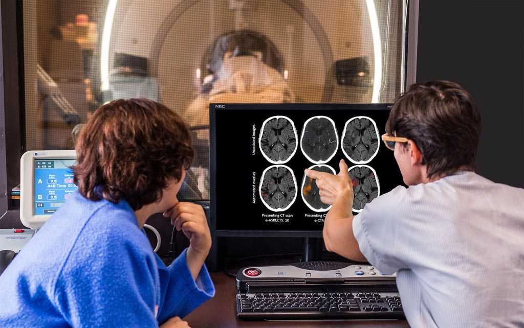
Image: The e-CTA software aids consistent mechanical thrombectomy patient selection (Photo courtesy of Brainomix).
A novel CT angiography (CTA) imaging solution improves inter-rater consistency and reliability, helping the selection of patients that can benefit the most from mechanical thrombectomy.
The Brainomix (Oxford, United Kingdom) e-CTA software is designed to provide fast, consistent, and fully automated calculation of the CTA collateral score (CTA-CS), using artificial intelligence (AI) and deep learning algorithms to analyze the images. In a clinical validation study, researchers at the University of Oxford (United Kingdom) categorized the degree of collateral flow in 98 patients, generating a CTA-CS score for each one. The results of the e-CTA software were then compared to a reference standard consensus opinion by from three expert neuroradiologists.
The agreement between individual neuroradiologists, both with and without the automated e-CTA reference was then quantified, and the effect of e-CTA on inter-rater reliability was assessed. The results demonstrated a high degree of agreement between the fully automated and objective e-CTA score and the consensus expert CTA-CS. The addition of an e-CTA assisted neuroradiological grading of collateral scores further reduced expert variability. CTA-CS and e-CTA scores correlated with the demonstrated deleterious effect of poor collateral blood flow on tissue survival. The study was published on June 19, 2019, in Cerebrovascular Disease.
“The importance of good collateral blood flow for patient outcome in acute stroke treatment is well recognized. However, even expert collateral scores commonly suffer from inter-rater error. e-CTA improved consistency between neuroradiologists when used as a decision support tool,” said George Harston, chief medical and innovation officer at Brainomix. “Even when used as a standalone tool, e-CTA produced similar scores compared to experts, opening up the opportunity for reliable collateral assessment for all patients, regardless of which hospital they attend.”
Timely restoration of cerebral blood flow using reperfusion therapy is the most effective maneuver for salvaging ischemic brain tissue that is not already infracted. Intravenous alteplase is first-line therapy, provided that treatment is initiated within 4.5 hours of clearly defined symptom onset. Mechanical thrombectomy is indicated for patients with acute ischemic stroke, due to a large artery occlusion in the anterior circulation, who can be treated within 24 hours of the time last known to be well, regardless of whether they receive intravenous alteplase for the same ischemic stroke event.
Related Links:
Brainomix
University of Oxford
Latest General/Advanced Imaging News
- AI Tool Offers Prognosis for Patients with Head and Neck Cancer
- New 3D Imaging System Addresses MRI, CT and Ultrasound Limitations
- AI-Based Tool Predicts Future Cardiovascular Events in Angina Patients
- AI-Based Tool Accelerates Detection of Kidney Cancer
- New Algorithm Dramatically Speeds Up Stroke Detection Scans
- 3D Scanning Approach Enables Ultra-Precise Brain Surgery
- AI Tool Improves Medical Imaging Process by 90%
- New Ultrasmall, Light-Sensitive Nanoparticles Could Serve as Contrast Agents
- AI Algorithm Accurately Predicts Pancreatic Cancer Metastasis Using Routine CT Images
- Cutting-Edge Angio-CT Solution Offers New Therapeutic Possibilities
- Extending CT Imaging Detects Hidden Blood Clots in Stroke Patients
- Groundbreaking AI Model Accurately Segments Liver Tumors from CT Scans
- New CT-Based Indicator Helps Predict Life-Threatening Postpartum Bleeding Cases
- CT Colonography Beats Stool DNA Testing for Colon Cancer Screening
- First-Of-Its-Kind Wearable Device Offers Revolutionary Alternative to CT Scans
- AI-Based CT Scan Analysis Predicts Early-Stage Kidney Damage Due to Cancer Treatments
Channels
Radiography
view channel
Routine Mammograms Could Predict Future Cardiovascular Disease in Women
Mammograms are widely used to screen for breast cancer, but they may also contain overlooked clues about cardiovascular health. Calcium deposits in the arteries of the breast signal stiffening blood vessels,... Read more
AI Detects Early Signs of Aging from Chest X-Rays
Chronological age does not always reflect how fast the body is truly aging, and current biological age tests often rely on DNA-based markers that may miss early organ-level decline. Detecting subtle, age-related... Read moreMRI
view channel
New Material Boosts MRI Image Quality
Magnetic resonance imaging (MRI) is a cornerstone of modern diagnostics, yet certain deep or anatomically complex tissues, including delicate structures of the eye and orbit, remain difficult to visualize clearly.... Read more
AI Model Reads and Diagnoses Brain MRI in Seconds
Brain MRI scans are critical for diagnosing strokes, hemorrhages, and other neurological disorders, but interpreting them can take hours or even days due to growing demand and limited specialist availability.... Read moreMRI Scan Breakthrough to Help Avoid Risky Invasive Tests for Heart Patients
Heart failure patients often require right heart catheterization to assess how severely their heart is struggling to pump blood, a procedure that involves inserting a tube into the heart to measure blood... Read more
MRI Scans Reveal Signature Patterns of Brain Activity to Predict Recovery from TBI
Recovery after traumatic brain injury (TBI) varies widely, with some patients regaining full function while others are left with lasting disabilities. Prognosis is especially difficult to assess in patients... Read moreUltrasound
view channel
Reusable Gel Pad Made from Tamarind Seed Could Transform Ultrasound Examinations
Ultrasound imaging depends on a conductive gel to eliminate air between the probe and the skin so sound waves can pass clearly into the body. While the imaging technology is fast, safe, and noninvasive,... Read more
AI Model Accurately Detects Placenta Accreta in Pregnancy Before Delivery
Placenta accreta spectrum (PAS) is a life-threatening pregnancy complication in which the placenta abnormally attaches to the uterine wall. The condition is a leading cause of maternal mortality and morbidity... Read moreNuclear Medicine
view channel
Radiopharmaceutical Molecule Marker to Improve Choice of Bladder Cancer Therapies
Targeted cancer therapies only work when tumor cells express the specific molecular structures they are designed to attack. In urothelial carcinoma, a common form of bladder cancer, the cell surface protein... Read more
Cancer “Flashlight” Shows Who Can Benefit from Targeted Treatments
Targeted cancer therapies can be highly effective, but only when a patient’s tumor expresses the specific protein the treatment is designed to attack. Determining this usually requires biopsies or advanced... Read moreImaging IT
view channel
New Google Cloud Medical Imaging Suite Makes Imaging Healthcare Data More Accessible
Medical imaging is a critical tool used to diagnose patients, and there are billions of medical images scanned globally each year. Imaging data accounts for about 90% of all healthcare data1 and, until... Read more
Global AI in Medical Diagnostics Market to Be Driven by Demand for Image Recognition in Radiology
The global artificial intelligence (AI) in medical diagnostics market is expanding with early disease detection being one of its key applications and image recognition becoming a compelling consumer proposition... Read moreIndustry News
view channel
Nuclear Medicine Set for Continued Growth Driven by Demand for Precision Diagnostics
Clinical imaging services face rising demand for precise molecular diagnostics and targeted radiopharmaceutical therapy as cancer and chronic disease rates climb. A new market analysis projects rapid expansion... Read more


 Guided Devices.jpg)


