AI Tool Accurately Assesses Ischemic Stroke Damage
|
By MedImaging International staff writers Posted on 04 Feb 2019 |

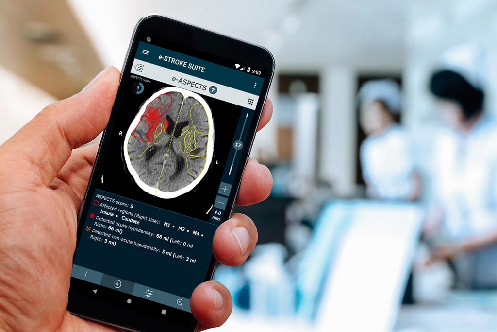
Image: The e-ASPECTS output as viewed on a mobile device (Photo courtesy of Brainomix).
An artificial intelligence (AI) imaging tool supports fast and consistent interpretation of non-contrast computerized tomography (CT) scan.
The Brainomix (Oxford, United Kingdom) e-ASPECTS tool is intended to assist clinicians in brain CT scan interpretation following an ischemic stroke by quantifying the volume of ischemia and grading it with the Alberta Stroke Program Early CT Score (ASPECTS) 10-point quantitative score. By reducing inter-reader variability in interpretation, e-ASPECTS enables a more standardized stroke diagnosis and facilitates fast, consistent treatment decisions, irrespective of physician experience or expertise.
Physicians can review e-ASPECTS results anywhere via picture archiving and communication systems (PACS), through the e-ASPECTS web browser user interface, or by viewing images sent to a clinician's smartphone via email. The wide availability ensures rapid sharing throughout the stroke team and allows faster, more informed decision-making. For users carrying out clinical research, the unique ischemia volume measurement feature in e-ASPECTS provides a fully automated estimate of the ischemic core size on non-contrast CT, even for hyper-acute cases.
A new study, published on October 27, 2018, in the Journal of NeuroInterventional Surgery, suggests that it may be reasonable to select patients for reperfusion therapy in hospitals without access to advanced imaging using the e-ASPECTS tool in combination with clinical criteria, and that e-ASPECTS is a reliable and valuable tool that can save valuable time by providing objective identification of ischemic injury, thus empowering clinicians in selecting patients suitable for mechanical thrombectomy, thrombolysis, endovascular treatment, or for decompressive craniotomy.
“This important study highlights the potential of the Brainomix e-ASPECTS support tool to simplify the selection of stroke patients for thrombectomy presenting in the late time window,” said George Harston, MD, chief medical and innovation officer of Brainomix and a consultant physician at Oxford University Hospitals. “The findings suggest that access to thrombectomy may be broadened to a wider population of patients, and without need for time consuming advanced imaging, which is often not readily available even in larger centers.”
Timely restoration of cerebral blood flow using reperfusion therapy is the most effective maneuver for salvaging ischemic brain tissue that is not already infarcted. For eligible patients with acute ischemic stroke, intravenous alteplase is first-line therapy, provided that treatment is initiated within 4.5 hours of onset. Mechanical thrombectomy is indicated for patients with acute ischemic stroke due to a large artery occlusion in the anterior circulation who can be treated within 24 hours of the time last known to be well, regardless of whether they receive intravenous alteplase for the same ischemic stroke event.
Related Links:
Brainomix
Latest Imaging IT News
- New Google Cloud Medical Imaging Suite Makes Imaging Healthcare Data More Accessible
- Global AI in Medical Diagnostics Market to Be Driven by Demand for Image Recognition in Radiology
- AI-Based Mammography Triage Software Helps Dramatically Improve Interpretation Process
- Artificial Intelligence (AI) Program Accurately Predicts Lung Cancer Risk from CT Images
- Image Management Platform Streamlines Treatment Plans
- AI-Based Technology for Ultrasound Image Analysis Receives FDA Approval
- AI Technology for Detecting Breast Cancer Receives CE Mark Approval
- Digital Pathology Software Improves Workflow Efficiency
- Patient-Centric Portal Facilitates Direct Imaging Access
- New Workstation Supports Customer-Driven Imaging Workflow
Channels
Radiography
view channel
Routine Mammograms Could Predict Future Cardiovascular Disease in Women
Mammograms are widely used to screen for breast cancer, but they may also contain overlooked clues about cardiovascular health. Calcium deposits in the arteries of the breast signal stiffening blood vessels,... Read more
AI Detects Early Signs of Aging from Chest X-Rays
Chronological age does not always reflect how fast the body is truly aging, and current biological age tests often rely on DNA-based markers that may miss early organ-level decline. Detecting subtle, age-related... Read moreMRI
view channel
MRI Scan Breakthrough to Help Avoid Risky Invasive Tests for Heart Patients
Heart failure patients often require right heart catheterization to assess how severely their heart is struggling to pump blood, a procedure that involves inserting a tube into the heart to measure blood... Read more
MRI Scans Reveal Signature Patterns of Brain Activity to Predict Recovery from TBI
Recovery after traumatic brain injury (TBI) varies widely, with some patients regaining full function while others are left with lasting disabilities. Prognosis is especially difficult to assess in patients... Read moreUltrasound
view channel
Portable Ultrasound Sensor to Enable Earlier Breast Cancer Detection
Breast cancer screening relies heavily on annual mammograms, but aggressive tumors can develop between scans, accounting for up to 30 percent of cases. These interval cancers are often diagnosed later,... Read more
Portable Imaging Scanner to Diagnose Lymphatic Disease in Real Time
Lymphatic disorders affect hundreds of millions of people worldwide and are linked to conditions ranging from limb swelling and organ dysfunction to birth defects and cancer-related complications.... Read more
Imaging Technique Generates Simultaneous 3D Color Images of Soft-Tissue Structure and Vasculature
Medical imaging tools often force clinicians to choose between speed, structural detail, and functional insight. Ultrasound is fast and affordable but typically limited to two-dimensional anatomy, while... Read moreNuclear Medicine
view channel
Radiopharmaceutical Molecule Marker to Improve Choice of Bladder Cancer Therapies
Targeted cancer therapies only work when tumor cells express the specific molecular structures they are designed to attack. In urothelial carcinoma, a common form of bladder cancer, the cell surface protein... Read more
Cancer “Flashlight” Shows Who Can Benefit from Targeted Treatments
Targeted cancer therapies can be highly effective, but only when a patient’s tumor expresses the specific protein the treatment is designed to attack. Determining this usually requires biopsies or advanced... Read moreImaging IT
view channel
New Google Cloud Medical Imaging Suite Makes Imaging Healthcare Data More Accessible
Medical imaging is a critical tool used to diagnose patients, and there are billions of medical images scanned globally each year. Imaging data accounts for about 90% of all healthcare data1 and, until... Read more
Global AI in Medical Diagnostics Market to Be Driven by Demand for Image Recognition in Radiology
The global artificial intelligence (AI) in medical diagnostics market is expanding with early disease detection being one of its key applications and image recognition becoming a compelling consumer proposition... Read moreIndustry News
view channel
Nuclear Medicine Set for Continued Growth Driven by Demand for Precision Diagnostics
Clinical imaging services face rising demand for precise molecular diagnostics and targeted radiopharmaceutical therapy as cancer and chronic disease rates climb. A new market analysis projects rapid expansion... Read more


