Wide Bore CT Enhances Oncologic Radiotherapy Treatment
|
By MedImaging International staff writers Posted on 15 Nov 2018 |

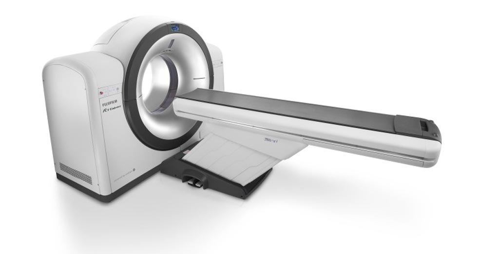
Image: An 85cm wide bore facilitates CT imaging and nuclear medicine (Photo courtesy of Fujifilm).
A novel 85cm wide bore computed tomography (CT) imaging unit offers enhanced and efficient CT radiotherapy treatment (RT) planning capabilities.
The Fujifilm (Tokyo, Japan) FCT Embrace is designed to improve accuracy throughout the entire oncology care cycle, by optimally matching the rotational arc of the linear accelerator (LINAC) with that of the wide bore, thus providing precise positioning options for both preliminary simulation and RT treatment planning. Oncology patients can be imaged in their optimal treatment position at the full clinical image quality afforded by 64 slice or 128 slice configurations. The FCT Embrace is comprised of just 4 major parts with 100 components that connect seamlessly using brushless PowerLink slip-ring technology, thus streamlining installation and maintenance.
For radiology and radiation oncology professionals who face logistical complexity while making high-stakes care decisions, the FCT Embrace also features a modular platform to seamlessly serve the needs of a wide range of medical specialties, spanning oncology, pediatrics, geriatrics, orthopedics, and emergency care. The 85-cm bore is one-size-fits-all, comfortably accommodating virtually any patient, from bariatric to pediatric; and procedure, from prone breast board to large biopsy needle placement.
Additionally, the bore aperture is conveniently suited to RT treatment planning software, easing the transition from radiology to oncology. Innovative 4D gating technology accounts for patient movement while breathing, allowing more targeted treatment options. The FCT Embrace also offers the industry’s widest table at 49 cm, minimizing the risk of falling and maximizing comfort for a wide range of patients, including bariatric patients weighing up to 300 kilograms.
“Our goal is to simplify the workflow of radiation oncology professionals. We are delivering on this promise with the FCT Embrace, a one-stop solution for the entire radiation therapy workflow offering CT imaging, simulation, treatment planning, and more,” said Alan Glenn, vice president of sales at Fujifilm. “Its sheer size at 85-cm bore optimally matches the rotational arc of the linear accelerator to offer easier and precise positioning options for simulation while providing an open patient experience. The modular platform maximizes synergies of care.”
“In 1936, we took our first steps in the development of X-ray film; and in 1983, we pioneered the first digitized radiography system in the world,” said Johann Fernando, PhD, Chief Operating Officer of Fujifilm Medical Systems USA. “Once again we are innovating with the launch of the FCT Embrace, a solution that provides the most slices ever seen on an 85cm bore system. Designed to improve radiation oncology care, this advanced solution boosts patient comfort and security by offering the widest tabletop currently available.”
Latest General/Advanced Imaging News
- AI-Powered Imaging System Improves Lung Cancer Diagnosis
- AI Model Significantly Enhances Low-Dose CT Capabilities
- Ultra-Low Dose CT Aids Pneumonia Diagnosis in Immunocompromised Patients
- AI Reduces CT Lung Cancer Screening Workload by Almost 80%
- Cutting-Edge Technology Combines Light and Sound for Real-Time Stroke Monitoring
- AI System Detects Subtle Changes in Series of Medical Images Over Time
- New CT Scan Technique to Improve Prognosis and Treatments for Head and Neck Cancers
- World’s First Mobile Whole-Body CT Scanner to Provide Diagnostics at POC
- Comprehensive CT Scans Could Identify Atherosclerosis Among Lung Cancer Patients
- AI Improves Detection of Colorectal Cancer on Routine Abdominopelvic CT Scans
- Super-Resolution Technology Enhances Clinical Bone Imaging to Predict Osteoporotic Fracture Risk
- AI-Powered Abdomen Map Enables Early Cancer Detection
- Deep Learning Model Detects Lung Tumors on CT
- AI Predicts Cardiovascular Risk from CT Scans
- Deep Learning Based Algorithms Improve Tumor Detection in PET/CT Scans
- New Technology Provides Coronary Artery Calcification Scoring on Ungated Chest CT Scans
Channels
Radiography
view channel
AI-Powered Imaging Technique Shows Promise in Evaluating Patients for PCI
Percutaneous coronary intervention (PCI), also known as coronary angioplasty, is a minimally invasive procedure where small metal tubes called stents are inserted into partially blocked coronary arteries... Read more
Higher Chest X-Ray Usage Catches Lung Cancer Earlier and Improves Survival
Lung cancer continues to be the leading cause of cancer-related deaths worldwide. While advanced technologies like CT scanners play a crucial role in detecting lung cancer, more accessible and affordable... Read moreMRI
view channel
Ultra-Powerful MRI Scans Enable Life-Changing Surgery in Treatment-Resistant Epileptic Patients
Approximately 360,000 individuals in the UK suffer from focal epilepsy, a condition in which seizures spread from one part of the brain. Around a third of these patients experience persistent seizures... Read more
AI-Powered MRI Technology Improves Parkinson’s Diagnoses
Current research shows that the accuracy of diagnosing Parkinson’s disease typically ranges from 55% to 78% within the first five years of assessment. This is partly due to the similarities shared by Parkinson’s... Read more
Biparametric MRI Combined with AI Enhances Detection of Clinically Significant Prostate Cancer
Artificial intelligence (AI) technologies are transforming the way medical images are analyzed, offering unprecedented capabilities in quantitatively extracting features that go beyond traditional visual... Read more
First-Of-Its-Kind AI-Driven Brain Imaging Platform to Better Guide Stroke Treatment Options
Each year, approximately 800,000 people in the U.S. experience strokes, with marginalized and minoritized groups being disproportionately affected. Strokes vary in terms of size and location within the... Read moreUltrasound
view channel
Smart Ultrasound-Activated Immune Cells Destroy Cancer Cells for Extended Periods
Chimeric antigen receptor (CAR) T-cell therapy has emerged as a highly promising cancer treatment, especially for bloodborne cancers like leukemia. This highly personalized therapy involves extracting... Read more
Tiny Magnetic Robot Takes 3D Scans from Deep Within Body
Colorectal cancer ranks as one of the leading causes of cancer-related mortality worldwide. However, when detected early, it is highly treatable. Now, a new minimally invasive technique could significantly... Read more
High Resolution Ultrasound Speeds Up Prostate Cancer Diagnosis
Each year, approximately one million prostate cancer biopsies are conducted across Europe, with similar numbers in the USA and around 100,000 in Canada. Most of these biopsies are performed using MRI images... Read more
World's First Wireless, Handheld, Whole-Body Ultrasound with Single PZT Transducer Makes Imaging More Accessible
Ultrasound devices play a vital role in the medical field, routinely used to examine the body's internal tissues and structures. While advancements have steadily improved ultrasound image quality and processing... Read moreNuclear Medicine
view channel
Novel PET Imaging Approach Offers Never-Before-Seen View of Neuroinflammation
COX-2, an enzyme that plays a key role in brain inflammation, can be significantly upregulated by inflammatory stimuli and neuroexcitation. Researchers suggest that COX-2 density in the brain could serve... Read more
Novel Radiotracer Identifies Biomarker for Triple-Negative Breast Cancer
Triple-negative breast cancer (TNBC), which represents 15-20% of all breast cancer cases, is one of the most aggressive subtypes, with a five-year survival rate of about 40%. Due to its significant heterogeneity... Read moreImaging IT
view channel
New Google Cloud Medical Imaging Suite Makes Imaging Healthcare Data More Accessible
Medical imaging is a critical tool used to diagnose patients, and there are billions of medical images scanned globally each year. Imaging data accounts for about 90% of all healthcare data1 and, until... Read more
Global AI in Medical Diagnostics Market to Be Driven by Demand for Image Recognition in Radiology
The global artificial intelligence (AI) in medical diagnostics market is expanding with early disease detection being one of its key applications and image recognition becoming a compelling consumer proposition... Read moreIndustry News
view channel
GE HealthCare and NVIDIA Collaboration to Reimagine Diagnostic Imaging
GE HealthCare (Chicago, IL, USA) has entered into a collaboration with NVIDIA (Santa Clara, CA, USA), expanding the existing relationship between the two companies to focus on pioneering innovation in... Read more
Patient-Specific 3D-Printed Phantoms Transform CT Imaging
New research has highlighted how anatomically precise, patient-specific 3D-printed phantoms are proving to be scalable, cost-effective, and efficient tools in the development of new CT scan algorithms... Read more
Siemens and Sectra Collaborate on Enhancing Radiology Workflows
Siemens Healthineers (Forchheim, Germany) and Sectra (Linköping, Sweden) have entered into a collaboration aimed at enhancing radiologists' diagnostic capabilities and, in turn, improving patient care... Read more


