Robot-Supported Angiography System Advances Hybrid ORs
|
By Simone Ciolek Posted on 28 Oct 2016 |

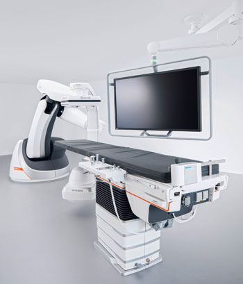
Image: The Artis Pheno robot-supported angiography system (Photo courtesy of Siemens Healthineers).
A novel angiography system offers personalized, minimally invasive surgery for multimorbid patients with changing disease patterns.
The Siemens Healthineers (Erlangen, Germany) Artis pheno provides patients with multiple health issues the option to undergo MIS and interventional radiology and interventional cardiology procedures, despite the additional risks associated with chronic disease. Artis can scan the body up to 15% faster (compared to prior systems Siemens Healthineers systems), and produce three-dimensional (3D) images that need less contrast agent; if the patient is sensitive to the contrast agent, Artis pheno can also support CO2 imaging of the extremities.
The C-arm is 13 centimeters wider and has a free inner diameter of 95.5 centimeters, offering more space for handling adipose patients and longer instrument use that can be used without added difficulty. The multi-tilt table is designed to accommodate patients weighing up to 280 kilograms, with edge of the table capable of tilting to stabilize the patient's blood pressure, for example, or to make breathing easier when necessary. The system can be fitted with a comprehensive range of optional software applications to deal with complex cases.
The robotic construction of the Siemens Healthineers table gives it a flexible isocenter, so that the angiography system can follow all table positions and provide imaging support for the patient's treatment, while representing the target area of the body from virtually any angle. Artis pheno recognizes the position of the tabletop at all times, and automatically aligns itself. Memory positions let the system move the C-arm out of the operating area quickly if necessary, giving the surgeon and team free access to the patient, and then move it back to exactly the same position again for further imaging.
A dedicated hygiene approach combines several factors: an antimicrobial coating prevents bacteria and viruses from multiplying on the system; the wiring is routed inside the system to prevent cables from becoming dirty and potentially transmitting bacteria; and seamless surfaces with no recesses, and spaces that are easy to access, make the system easier to clean. And because the system is floor-mounted, it is easier to install in the OR, and the sterile airflow from the ceiling is interrupted during imaging only by the flat-panel detector.
“We see a high number of multimorbid patients with impaired kidney function in the angio suite,” said Professor Frank Wacker, MD, director of the institute for diagnostic and interventional radiology at Hanover Medical School (Germany). “Shorter scan times help reduce the amount of iodinated contrast agent during 3D angiography in the thorax and abdomen by up to fifteen percent.”
Additional optional application packages include software for extensive spinal fusion procedures using screws or needles; screw paths can be precisely planned, and an automatic path alignment function automatically aligns the C-arm to follow them. A laser integrated in the image detector shows the surgeon the planned path, which helps improve both accuracy and speed in the OR. A number of applications support tumor transarterial chemoembolization (TACE), rendering arterial vessels visible, with graphic overlaying of the selected vessel paths with the real-time X-ray images.
Related Links:
Siemens Healthineers
Hanover Medical School
Latest General/Advanced Imaging News
- AI-Based CT Scan Analysis Predicts Early-Stage Kidney Damage Due to Cancer Treatments
- CT-Based Deep Learning-Driven Tool to Enhance Liver Cancer Diagnosis
- AI-Powered Imaging System Improves Lung Cancer Diagnosis
- AI Model Significantly Enhances Low-Dose CT Capabilities
- Ultra-Low Dose CT Aids Pneumonia Diagnosis in Immunocompromised Patients
- AI Reduces CT Lung Cancer Screening Workload by Almost 80%
- Cutting-Edge Technology Combines Light and Sound for Real-Time Stroke Monitoring
- AI System Detects Subtle Changes in Series of Medical Images Over Time
- New CT Scan Technique to Improve Prognosis and Treatments for Head and Neck Cancers
- World’s First Mobile Whole-Body CT Scanner to Provide Diagnostics at POC
- Comprehensive CT Scans Could Identify Atherosclerosis Among Lung Cancer Patients
- AI Improves Detection of Colorectal Cancer on Routine Abdominopelvic CT Scans
- Super-Resolution Technology Enhances Clinical Bone Imaging to Predict Osteoporotic Fracture Risk
- AI-Powered Abdomen Map Enables Early Cancer Detection
- Deep Learning Model Detects Lung Tumors on CT
- AI Predicts Cardiovascular Risk from CT Scans
Channels
Radiography
view channel
AI Improves Early Detection of Interval Breast Cancers
Interval breast cancers, which occur between routine screenings, are easier to treat when detected earlier. Early detection can reduce the need for aggressive treatments and improve the chances of better outcomes.... Read more
World's Largest Class Single Crystal Diamond Radiation Detector Opens New Possibilities for Diagnostic Imaging
Diamonds possess ideal physical properties for radiation detection, such as exceptional thermal and chemical stability along with a quick response time. Made of carbon with an atomic number of six, diamonds... Read moreMRI
view channel
Cutting-Edge MRI Technology to Revolutionize Diagnosis of Common Heart Problem
Aortic stenosis is a common and potentially life-threatening heart condition. It occurs when the aortic valve, which regulates blood flow from the heart to the rest of the body, becomes stiff and narrow.... Read more
New MRI Technique Reveals True Heart Age to Prevent Attacks and Strokes
Heart disease remains one of the leading causes of death worldwide. Individuals with conditions such as diabetes or obesity often experience accelerated aging of their hearts, sometimes by decades.... Read more
AI Tool Predicts Relapse of Pediatric Brain Cancer from Brain MRI Scans
Many pediatric gliomas are treatable with surgery alone, but relapses can be catastrophic. Predicting which patients are at risk for recurrence remains challenging, leading to frequent follow-ups with... Read more
AI Tool Tracks Effectiveness of Multiple Sclerosis Treatments Using Brain MRI Scans
Multiple sclerosis (MS) is a condition in which the immune system attacks the brain and spinal cord, leading to impairments in movement, sensation, and cognition. Magnetic Resonance Imaging (MRI) markers... Read moreUltrasound
view channel.jpeg)
AI-Powered Lung Ultrasound Outperforms Human Experts in Tuberculosis Diagnosis
Despite global declines in tuberculosis (TB) rates in previous years, the incidence of TB rose by 4.6% from 2020 to 2023. Early screening and rapid diagnosis are essential elements of the World Health... Read more
AI Identifies Heart Valve Disease from Common Imaging Test
Tricuspid regurgitation is a condition where the heart's tricuspid valve does not close completely during contraction, leading to backward blood flow, which can result in heart failure. A new artificial... Read moreNuclear Medicine
view channel
Novel Radiolabeled Antibody Improves Diagnosis and Treatment of Solid Tumors
Interleukin-13 receptor α-2 (IL13Rα2) is a cell surface receptor commonly found in solid tumors such as glioblastoma, melanoma, and breast cancer. It is minimally expressed in normal tissues, making it... Read more
Novel PET Imaging Approach Offers Never-Before-Seen View of Neuroinflammation
COX-2, an enzyme that plays a key role in brain inflammation, can be significantly upregulated by inflammatory stimuli and neuroexcitation. Researchers suggest that COX-2 density in the brain could serve... Read moreImaging IT
view channel
New Google Cloud Medical Imaging Suite Makes Imaging Healthcare Data More Accessible
Medical imaging is a critical tool used to diagnose patients, and there are billions of medical images scanned globally each year. Imaging data accounts for about 90% of all healthcare data1 and, until... Read more
Global AI in Medical Diagnostics Market to Be Driven by Demand for Image Recognition in Radiology
The global artificial intelligence (AI) in medical diagnostics market is expanding with early disease detection being one of its key applications and image recognition becoming a compelling consumer proposition... Read moreIndustry News
view channel
GE HealthCare and NVIDIA Collaboration to Reimagine Diagnostic Imaging
GE HealthCare (Chicago, IL, USA) has entered into a collaboration with NVIDIA (Santa Clara, CA, USA), expanding the existing relationship between the two companies to focus on pioneering innovation in... Read more
Patient-Specific 3D-Printed Phantoms Transform CT Imaging
New research has highlighted how anatomically precise, patient-specific 3D-printed phantoms are proving to be scalable, cost-effective, and efficient tools in the development of new CT scan algorithms... Read more
Siemens and Sectra Collaborate on Enhancing Radiology Workflows
Siemens Healthineers (Forchheim, Germany) and Sectra (Linköping, Sweden) have entered into a collaboration aimed at enhancing radiologists' diagnostic capabilities and, in turn, improving patient care... Read more


