Dynamic CMOS Flat X-Ray Detectors Designed for Interventional Procedures
|
By MedImaging International staff writers Posted on 11 Dec 2014 |

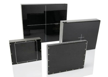
Image: The Xineos range of real time, low dose complementary metal-oxide semiconductor (CMOS) flat X-ray detectors (Photo courtesy of Teledyne Dalsa).
Radiation-hard detectors with improved dose performance and extended dynamic range provides switchable saturation dose, low dissipation power and built-in gain, offset and defect correction, a range of radiation-hard detectors are versatile, effective, and easy to integrate.
Teledyne Dalsa (Waterloo, ON, Canada), a Teledyne Technologies company and developer of digital X-ray image sensing technology, presented four new dynamic detectors at the Radiological Society of North America (RSNA) 2014, November 30 to December 5, 2014, in Chicago (IL, USA). The advanced detectors with fields-of-view (FOV) of 20 x 22 cm, 22 x 22 cm and 30 x 30 cm, together with earlier models with dimensions of 13 x 13 cm and 15 x 15 cm, complete the Xineos range of real time, low dose complementary metal-oxide semiconductor (CMOS) flat X-ray detectors. Xineos detectors are targeted at interventional procedures and are well-positioned to replace the incumbent imaging technology of thin-film transistor (TFT) flat detectors and image intensifiers of up to 30.48 cm in size.
The new Xineos models, including the 2222HS, 3030HS, 2022HR, and 3030HR, utilize Teledyne Dalsa’s sixth-generation proprietary radiation-hard CMOS active pixel design and are the industry’s first flat detectors to not only surpass the low-dose performance of image intensifiers but also combine this industry benchmark with the form factor advantages of traditional amorphous silicon flat panels. Offering switchable saturation dose, low dissipation power and built-in gain, offset and defect correction, Xineos detectors are versatile, effective, and easy to integrate.
The high speed Xineos-HS detectors, featuring 152 μm pixel size, are designed to meet the demanding needs of both surgical and cardiovascular procedures by combining high dynamic range with unsurpassed signal-to-noise performance and real-time imaging at the lowest exposure levels in full resolution at up to 90 fps. Additionally, the detectors with square FOV of 22 x 22 cm2 are the largest commercially available 9-inch flat detectors to benefit spinal surgery.
Xineos-HR is the high resolution line of detectors, featuring pixel size of 99 micrometers, designed to meet the versatile needs of clinical, scientific and industrial applications. These detectors are capable of 30 fps real-time imaging at full resolution of up to 5 Mpixels or 9 Mpixels for the 9-inch or 12-inch detector sizes, respectively. Flexible zoom modes can be used to capture regions-of-interest at 120 fps or even faster.
“Xineos CMOS detectors enable improvement throughout the entire healthcare chain—in diagnostics, in patient care, and in their return on investment,” said Dr. Mila Heeman, senior marketing manager at Teledyne Dalsa. “Our detectors allow physicians to expand the boundaries of imaging.”
Teledyne DALSA exhibited the following CMOS and charged coupled detector (CCD) X-ray detectors at RSNA: (1) Xineos-1313 and Xineos CMOS flat detectors provide low dose diagnostic image quality at full resolution and in real time. There are two versions: high dynamic range and high sensitivity. (2) The Xineos-1515 features streamlined switchable saturation dose for combining high dynamic range and high sensitivity in a single device. (3) Argus scanning CCD-TDI X-ray detector platform is designed for mammography, general radiography, and dental panoramic and cephalometry applications. (4) The Helios isoptimized for mobile applications, and features a large CMOS active imaging area of 20 x 25 cm2 (8-inch x10-inch) while maintaining excellent image resolution with a pixel size of only 96 micrometers. (5) The Shad-o-Box 6K HS is a high resolution CMOS detector with 50 μm pixel pitch and dimensions of 11 x 14 cm targeted at biopsy analysis, and other scientific and industrial applications. (6) Lastly, the Rad-icon 1520 is a new high resolution large area CMOS detector featuring high speed low dose seamless switchable saturation dose and 99 μm pixel in 15 x 20 cm dimensions.
Related Links:
Teledyne Dalsa
Latest Radiography News
- Machine Learning Algorithm Identifies Cardiovascular Risk from Routine Bone Density Scans
- AI Improves Early Detection of Interval Breast Cancers
- World's Largest Class Single Crystal Diamond Radiation Detector Opens New Possibilities for Diagnostic Imaging
- AI-Powered Imaging Technique Shows Promise in Evaluating Patients for PCI
- Higher Chest X-Ray Usage Catches Lung Cancer Earlier and Improves Survival
- AI-Powered Mammograms Predict Cardiovascular Risk
- Generative AI Model Significantly Reduces Chest X-Ray Reading Time
- AI-Powered Mammography Screening Boosts Cancer Detection in Single-Reader Settings
- Photon Counting Detectors Promise Fast Color X-Ray Images
- AI Can Flag Mammograms for Supplemental MRI
- 3D CT Imaging from Single X-Ray Projection Reduces Radiation Exposure
- AI Method Accurately Predicts Breast Cancer Risk by Analyzing Multiple Mammograms
- Printable Organic X-Ray Sensors Could Transform Treatment for Cancer Patients
- Highly Sensitive, Foldable Detector to Make X-Rays Safer
- Novel Breast Cancer Screening Technology Could Offer Superior Alternative to Mammogram
- Artificial Intelligence Accurately Predicts Breast Cancer Years Before Diagnosis
Channels
MRI
view channel
MRI to Replace Painful Spinal Tap for Faster MS Diagnosis
Multiple sclerosis (MS) is a challenging neurological condition to diagnose due to its wide array of symptoms, with not all patients experiencing the same symptoms or at the same intensity, and the disease... Read more
MRI Scans Can Identify Cardiovascular Disease Ten Years in Advance
Cardiovascular disease encompasses various conditions that narrow or block blood vessels, such as heart attacks, strokes, and heart failure. While some individuals are genetically predisposed, lifestyle... Read more
Simple Brain Scan Diagnoses Parkinson's Disease Years Before It Becomes Untreatable
Parkinson's disease (PD) remains a challenging condition to treat, with no known cure. Though therapies have improved over time, and ongoing research focuses on methods to slow or alter the disease’s progression,... Read moreUltrasound
view channel
New Incision-Free Technique Halts Growth of Debilitating Brain Lesions
Cerebral cavernous malformations (CCMs), also known as cavernomas, are abnormal clusters of blood vessels that can grow in the brain, spinal cord, or other parts of the body. While most cases remain asymptomatic,... Read more.jpeg)
AI-Powered Lung Ultrasound Outperforms Human Experts in Tuberculosis Diagnosis
Despite global declines in tuberculosis (TB) rates in previous years, the incidence of TB rose by 4.6% from 2020 to 2023. Early screening and rapid diagnosis are essential elements of the World Health... Read moreNuclear Medicine
view channel
New Imaging Approach Could Reduce Need for Biopsies to Monitor Prostate Cancer
Prostate cancer is the second leading cause of cancer-related death among men in the United States. However, the majority of older men diagnosed with prostate cancer have slow-growing, low-risk forms of... Read more
Novel Radiolabeled Antibody Improves Diagnosis and Treatment of Solid Tumors
Interleukin-13 receptor α-2 (IL13Rα2) is a cell surface receptor commonly found in solid tumors such as glioblastoma, melanoma, and breast cancer. It is minimally expressed in normal tissues, making it... Read moreGeneral/Advanced Imaging
view channel
First-Of-Its-Kind Wearable Device Offers Revolutionary Alternative to CT Scans
Currently, patients with conditions such as heart failure, pneumonia, or respiratory distress often require multiple imaging procedures that are intermittent, disruptive, and involve high levels of radiation.... Read more
AI-Based CT Scan Analysis Predicts Early-Stage Kidney Damage Due to Cancer Treatments
Radioligand therapy, a form of targeted nuclear medicine, has recently gained attention for its potential in treating specific types of tumors. However, one of the potential side effects of this therapy... Read moreImaging IT
view channel
New Google Cloud Medical Imaging Suite Makes Imaging Healthcare Data More Accessible
Medical imaging is a critical tool used to diagnose patients, and there are billions of medical images scanned globally each year. Imaging data accounts for about 90% of all healthcare data1 and, until... Read more
Global AI in Medical Diagnostics Market to Be Driven by Demand for Image Recognition in Radiology
The global artificial intelligence (AI) in medical diagnostics market is expanding with early disease detection being one of its key applications and image recognition becoming a compelling consumer proposition... Read moreIndustry News
view channel
GE HealthCare and NVIDIA Collaboration to Reimagine Diagnostic Imaging
GE HealthCare (Chicago, IL, USA) has entered into a collaboration with NVIDIA (Santa Clara, CA, USA), expanding the existing relationship between the two companies to focus on pioneering innovation in... Read more
Patient-Specific 3D-Printed Phantoms Transform CT Imaging
New research has highlighted how anatomically precise, patient-specific 3D-printed phantoms are proving to be scalable, cost-effective, and efficient tools in the development of new CT scan algorithms... Read more
Siemens and Sectra Collaborate on Enhancing Radiology Workflows
Siemens Healthineers (Forchheim, Germany) and Sectra (Linköping, Sweden) have entered into a collaboration aimed at enhancing radiologists' diagnostic capabilities and, in turn, improving patient care... Read more


