Four Imaging Approaches Can Differentiate Malignant and Benign Breast Tumors
|
By MedImaging International staff writers Posted on 10 Jul 2014 |

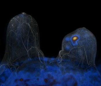
Image: A PET/MRI scanner visualizes breast cancer in this fused PET/MR image (Photo courtesy of Medscape).
Imaging breast tumors using four approaches together, can better distinguish malignant breast tumors from those that are benign, compared with imaging using fewer approaches, and this may help avoid repeat breast biopsies.
The findings were published online June 24, 2014, in Clinical Cancer Research, a journal of the American Association for Cancer Research. “By assessing many functional processes involved in cancer development, a multiparameter PET/MRI [positron emission tomography/magnetic resonance imaging] of the breast allows for a better differentiation of benign and malignant breast tumors than currently used dynamic contrast-enhanced MRI [DCE-MRI)] by itself. Therefore, unnecessary breast biopsies can be avoided,” said Katja Pinker, MD, associate professor of radiology in the department of biomedical imaging and image-guided therapy at the Medical University of Vienna (Austria).
The new imaging technique, called multiparametric (MP) 18FDG (fluorodeoxyglucose) PET-MRI, which used four imaging approaches, was 96% accurate in distinguishing malignant breast tumors from those that were benign, and provided better results than combinations of two or three imaging approaches. The study estimates that this technique can cut unnecessary breast biopsies recommended by the typically used imaging modality, the DCE-MRI, by 50%.
“DCE-MRI is a very sensitive test for the detection of breast tumors, but is limited in visualizing the functional properties cancer cells acquire during development. Therefore, there is still room for improvement,” explained Dr. Pinker. “PET/MRI mirrors cancer biology and allows accurate diagnosis of breast cancer without a biopsy. Additionally, the more accurately we understand a tumor’s biology, the better we can tailor therapy to each breast cancer patient’s individual needs. Provided that a hospital is equipped with a PET/CT [computed tomography] and an MRI scanner or a combined PET/MRI, the technique we have described can be immediately implemented in clinics,” said Dr. Pinker.
The investigators recruited 76 patients to the study who had suspicious or inconclusive findings from a mammography or a breast ultrasonography. They performed a MP18FDG PET/MRI on all the patients. Furthermore, all patients’ breast tumor biopsies were evaluated by histopathology.
To determine the combination of imaging parameters that yielded the most accurate results, Dr. Pinker and colleagues combined the imaging data from two parameters, three parameters, and all four parameters. All two-parameter and three-parameter evaluations included DCE-MRI. All findings were compared with histopathology diagnosis to evaluate which combination was most efficient in making an accurate diagnosis. Of the 76 tumors, based on histopathology, 53 were malignant and 23 were benign.
The researchers found that none of the two- or three-parameter combinations reached the same level of sensitivity and specificity as the four-parameter method, which had an appropriate use criteria (AUC) of 0.935. (An AUC of 0.9 to 1 means the modality is excellent, and an AUC of 0.5 means the method is worthless.) “Performing a combined PET-MRI is currently less cost-effective than existing breast imaging methods,” said Dr. Pinker. “However, a significant reduction in unnecessary breast biopsies by using this combined method may improve the cost-effectiveness.”
MP18FDG PET-MRI allowed tumor imaging by four parameters: DCE-MRI, diffusion-weighted imaging (DW), three-dimensional (3D) 1H-magnetic resonance spectroscopic imaging (MRSI), and 18FDG-PET.
Related Links:
Medical University of Vienna
Latest Nuclear Medicine News
- Novel Radiolabeled Antibody Improves Diagnosis and Treatment of Solid Tumors
- Novel PET Imaging Approach Offers Never-Before-Seen View of Neuroinflammation
- Novel Radiotracer Identifies Biomarker for Triple-Negative Breast Cancer
- Innovative PET Imaging Technique to Help Diagnose Neurodegeneration
- New Molecular Imaging Test to Improve Lung Cancer Diagnosis
- Novel PET Technique Visualizes Spinal Cord Injuries to Predict Recovery
- Next-Gen Tau Radiotracers Outperform FDA-Approved Imaging Agents in Detecting Alzheimer’s
- Breakthrough Method Detects Inflammation in Body Using PET Imaging
- Advanced Imaging Reveals Hidden Metastases in High-Risk Prostate Cancer Patients
- Combining Advanced Imaging Technologies Offers Breakthrough in Glioblastoma Treatment
- New Molecular Imaging Agent Accurately Identifies Crucial Cancer Biomarker
- New Scans Light Up Aggressive Tumors for Better Treatment
- AI Stroke Brain Scan Readings Twice as Accurate as Current Method
- AI Analysis of PET/CT Images Predicts Side Effects of Immunotherapy in Lung Cancer
- New Imaging Agent to Drive Step-Change for Brain Cancer Imaging
- Portable PET Scanner to Detect Earliest Stages of Alzheimer’s Disease
Channels
Radiography
view channel
AI Improves Early Detection of Interval Breast Cancers
Interval breast cancers, which occur between routine screenings, are easier to treat when detected earlier. Early detection can reduce the need for aggressive treatments and improve the chances of better outcomes.... Read more
World's Largest Class Single Crystal Diamond Radiation Detector Opens New Possibilities for Diagnostic Imaging
Diamonds possess ideal physical properties for radiation detection, such as exceptional thermal and chemical stability along with a quick response time. Made of carbon with an atomic number of six, diamonds... Read moreUltrasound
view channel.jpeg)
AI-Powered Lung Ultrasound Outperforms Human Experts in Tuberculosis Diagnosis
Despite global declines in tuberculosis (TB) rates in previous years, the incidence of TB rose by 4.6% from 2020 to 2023. Early screening and rapid diagnosis are essential elements of the World Health... Read more
AI Identifies Heart Valve Disease from Common Imaging Test
Tricuspid regurgitation is a condition where the heart's tricuspid valve does not close completely during contraction, leading to backward blood flow, which can result in heart failure. A new artificial... Read moreNuclear Medicine
view channel
Novel Radiolabeled Antibody Improves Diagnosis and Treatment of Solid Tumors
Interleukin-13 receptor α-2 (IL13Rα2) is a cell surface receptor commonly found in solid tumors such as glioblastoma, melanoma, and breast cancer. It is minimally expressed in normal tissues, making it... Read more
Novel PET Imaging Approach Offers Never-Before-Seen View of Neuroinflammation
COX-2, an enzyme that plays a key role in brain inflammation, can be significantly upregulated by inflammatory stimuli and neuroexcitation. Researchers suggest that COX-2 density in the brain could serve... Read moreGeneral/Advanced Imaging
view channel
CT-Based Deep Learning-Driven Tool to Enhance Liver Cancer Diagnosis
Medical imaging, such as computed tomography (CT) scans, plays a crucial role in oncology, offering essential data for cancer detection, treatment planning, and monitoring of response to therapies.... Read more
AI-Powered Imaging System Improves Lung Cancer Diagnosis
Given the need to detect lung cancer at earlier stages, there is an increasing need for a definitive diagnostic pathway for patients with suspicious pulmonary nodules. However, obtaining tissue samples... Read moreImaging IT
view channel
New Google Cloud Medical Imaging Suite Makes Imaging Healthcare Data More Accessible
Medical imaging is a critical tool used to diagnose patients, and there are billions of medical images scanned globally each year. Imaging data accounts for about 90% of all healthcare data1 and, until... Read more
Global AI in Medical Diagnostics Market to Be Driven by Demand for Image Recognition in Radiology
The global artificial intelligence (AI) in medical diagnostics market is expanding with early disease detection being one of its key applications and image recognition becoming a compelling consumer proposition... Read moreIndustry News
view channel
GE HealthCare and NVIDIA Collaboration to Reimagine Diagnostic Imaging
GE HealthCare (Chicago, IL, USA) has entered into a collaboration with NVIDIA (Santa Clara, CA, USA), expanding the existing relationship between the two companies to focus on pioneering innovation in... Read more
Patient-Specific 3D-Printed Phantoms Transform CT Imaging
New research has highlighted how anatomically precise, patient-specific 3D-printed phantoms are proving to be scalable, cost-effective, and efficient tools in the development of new CT scan algorithms... Read more
Siemens and Sectra Collaborate on Enhancing Radiology Workflows
Siemens Healthineers (Forchheim, Germany) and Sectra (Linköping, Sweden) have entered into a collaboration aimed at enhancing radiologists' diagnostic capabilities and, in turn, improving patient care... Read more


 Guided Devices.jpg)


