Digital Radiofluoroscopy Table System Receives FDA Clearance
|
By MedImaging International staff writers Posted on 08 May 2014 |
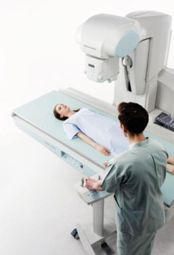
Image: Sonialvision G4 universal digital radiofluoroscopy (R/F) table system (Photo courtesy of Shimadzu Medical Systems).
A universal digital radiofluoroscopy (R/F) table system is designed to perform a wide range of examinations, including endoscopy, angiography, video fluoroscopy for gastrointestinal series and swallowing exams, orthopedic exams, and general radiography.
Shimadzu Medical Systems, USA (Torrance, CA, USA;) has announced that the Sonialvision G4 universal digital R/F table system has received 510(k) clearance from the US Food and Drug Administration (FDA). The system expands Shimadzu’s product range with the addition of the advanced system that has received strong acceptance in Europe and Asia.
The technology acquires high image quality images rapidly and uses low radiation dose levels. Its new ergonomic design is patient safety- and hygiene-focused, accommodates all sizes of ambulatory and stretcher/wheelchair confined patients, and offers workflow-efficiency features to help improve technologist productivity.
The system combines a 17-inch x 17-inch flat panel detector (FPD) with Shimadzu’s next- generation digital imaging platform. Its 139 μm ultra-small pixel pitch and high contrast maximizes the FPD performance. Its easily switchable large-to-small fields-of-view cover a range of imaging from finger tips to the entire chest or abdomen. Furthermore, the wide 202.5 cm movement range of the Sonialvision G4 can capture images of the complete body without moving the patient.
Various real-time digital filtering processes effectively suppress halation near skin surfaces and around shadows where organs overlap. Smoother imaging is achieved with no delay or image lags by applying optimal digital filtering and extracting noise components. Used in long spine and limb imaging, Shimadzu’s Slot Beam functionality is available as an advanced application option. With its lower dose profile, the Slot Beam is an excellent choice for pediatrics.
The Sonialvision G4 is equipped with a wealth of functions to facilitate urological exams. The edge of the imaging range can be positioned as close as 9.5 cm from the head end of the table, given a 17-inch field-of-view, making it easy to perform procedures using an optical endoscope. It is also possible to tilt the table without changing an observation position. A specialized collimation function helps reduce dose.
An oblique projection function prevents overlapping organs when performing gastrointestinal examinations. Fluoroscopic images can be saved in Digital Imaging and Communications in Medicine (DICOM) format. Patients in wheelchairs can be examined easily by extending the imaging chain, which has a maximum 15 cm per second quick movement.
The Sonialvision G4 supports angiography procedures with its streamlined design, and its large field of view enables digital subtraction angiography (DSA) to be used for exams ranging from the hepatic artery to the entire lower extremities. Special features for every type of exam the system performs help to minimize radiation dose exposure levels to the patient. A table that can support a patient weighing up to 317.5 kg in the horizontal position for bariatric procedures is designed for easy cleaning and integrates safety features to facilitate safe access and egress by patients.
This new system design of the G4 also provides productive and comfortable workflow to optimize convenience and operability for the technologists who use it. Its high-speed dual-processor can simultaneously resolve images, register patient data, and perform other functions rapidly without interfering with the examination being performed. The number of operating steps a technologist needs to perform during an examination is substantially reduced because the R/F table, X-ray generator, and digital processor are linked.
Related Links:
Shimadzu Medical Systems USA
Latest Radiography News
- Novel Breast Cancer Screening Technology Could Offer Superior Alternative to Mammogram
- Artificial Intelligence Accurately Predicts Breast Cancer Years Before Diagnosis
- AI-Powered Chest X-Ray Detects Pulmonary Nodules Three Years Before Lung Cancer Symptoms
- AI Model Identifies Vertebral Compression Fractures in Chest Radiographs
- Advanced 3D Mammography Detects More Breast Cancers
- AI X-Ray Diagnostic Tool Offers Rapid Pediatric Fracture Detection
- AI-Powered Chest X-Ray Analysis Shows Promise in Clinical Practice
- AI-Based Algorithm Improves Accuracy of Breast Cancer Diagnoses
- Groundbreaking X-Ray Imaging Technique Could Improve Medical Diagnostics
- Innovative X-Ray Technique Captures Human Heart with Unprecedented Detail
- Cutting-Edge Technology Enhances Chest X-Ray Classification for Superior Patient Outcomes
- AI Model Accurately Estimates Lung Function Using Chest X-Rays
- High-Powered Motorized Mobile C-Arm Delivers State-Of-The-Art Images for Challenging Procedures
- Injury Prediction Rule Reduces Radiographic Imaging Exposure in Children
- AI Detects More Breast Cancers with Fewer False Positives
- AI-Powered Portable Thermal Imaging Solution Could Complement Mammography for Breast Cancer Screening
Channels
MRI
view channel
Groundbreaking AI-Powered Software Significantly Enhances Brain MRI
Contrast-enhanced magnetic resonance imaging (MRI) utilizes contrast agents to illuminate specific tissues or abnormalities, leading to improved visualization and more comprehensive information.... Read more.jpeg)
MRI Predicts Patient Outcomes and Tumor Recurrence in Rectal Cancer Patients
Colorectal cancer is on the rise among younger adults—those under 50—while it has been declining in older populations. It is estimated that approximately 1 in 23 men and 1 in 25 women will be diagnosed... Read more
Portable MRI System Dramatically Cuts Time-To-Scan vs. Conventional MRI in Stroke Patients
Imaging of the brain is crucial for the management of acute stroke and transient ischemic attack. Key roles of imaging include confirming the diagnosis, determining treatment eligibility, indicating the... Read moreUltrasound
view channel
Ultrasound Can Identify Sources of Brain-Related Issues and Disorders Before Treatment
For many years, healthcare professionals worldwide have relied on ultrasound to monitor the growth of unborn infants and evaluate the health of internal organs. However, ultrasound technology, once primarily... Read more
New Guideline on Handling Endobronchial Ultrasound Transbronchial Needle Samples
Endobronchial ultrasound-guided transbronchial needle aspiration (EBUS-TBNA) has become the standard procedure for the initial diagnosis and staging of lung cancer; however, there is limited guidance on... Read more
Groundbreaking Ultrasound-Guided Needle Insertion System Improves Medical Procedures
Ultrasound-guided neuraxial procedures have traditionally been hampered by technical challenges, such as the need for three hands to manage the needle, probe, and syringe simultaneously, steep needle angles... Read moreNuclear Medicine
view channel
PET Software Enhances Diagnosis and Monitoring of Alzheimer's Disease
Alzheimer’s disease is marked by the buildup of beta-amyloid plaques and tau protein tangles in the brain. These deposits of beta-amyloid and tau appear in various brain regions at differing rates as the brain ages.... Read more.jpg)
New Photon-Counting CT Technique Diagnoses Osteoarthritis Before Symptoms Develop
X-ray imaging has evolved significantly since its introduction in 1895. This technique is known for capturing images quickly, making it ideal for emergency situations; however, it does not provide adequate... Read moreGeneral/Advanced Imaging
view channel
Low-Dose CT Screening for Lung Cancer Can Benefit Heavy Smokers
Lung cancer is often diagnosed at a late stage, with only about one-fifth to one-sixth of patients surviving five years after diagnosis. A new report now suggests that low-dose computed tomography (CT)... Read more![Image: A kidney showing positive [89Zr]Zr-girentuximab PET and histologically confirmed clear-cell renal cell carcinoma (Photo courtesy of Dr. Brian Shuch/UCLA Health) Image: A kidney showing positive [89Zr]Zr-girentuximab PET and histologically confirmed clear-cell renal cell carcinoma (Photo courtesy of Dr. Brian Shuch/UCLA Health)](https://globetechcdn.com/mobile_medicalimaging/images/stories/articles/article_images/2024-10-04/ca9scan.jpg)
Non-Invasive Imaging Technique Accurately Detects Aggressive Kidney Cancer
Kidney cancers, known as renal cell carcinomas, account for 90% of solid kidney tumors, with over 81,000 new cases diagnosed annually in the United States. Among the various types, clear-cell renal cell... Read more
AI Algorithm Reduces Unnecessary Radiation Exposure in Traumatic Neuroradiological CT Scans
Traumatic neuroradiological emergencies encompass conditions that require immediate and accurate diagnosis for effective treatment and optimal patient outcomes. These emergencies can include injuries to... Read more
New Solution Enhances AI-Based Quality Control and Diagnosis in Medical Imaging
Medical image data makes up 80% of clinical data, and artificial intelligence (AI) offers significant potential to unlock its full value, which is crucial for clinical diagnosis, decision-making, and disease... Read moreImaging IT
view channel
New Google Cloud Medical Imaging Suite Makes Imaging Healthcare Data More Accessible
Medical imaging is a critical tool used to diagnose patients, and there are billions of medical images scanned globally each year. Imaging data accounts for about 90% of all healthcare data1 and, until... Read more
Global AI in Medical Diagnostics Market to Be Driven by Demand for Image Recognition in Radiology
The global artificial intelligence (AI) in medical diagnostics market is expanding with early disease detection being one of its key applications and image recognition becoming a compelling consumer proposition... Read moreIndustry News
view channel.jpeg)
Philips and Medtronic Partner on Stroke Care
A stroke is typically an acute incident primarily caused by a blockage in a brain blood vessel, which disrupts the adequate blood supply to brain tissue and results in the permanent loss of brain cells.... Read more
Siemens and Medtronic Enter into Global Partnership for Advancing Spine Care Imaging Technologies
A new global partnership aims to explore opportunities to further expand access to advanced pre-and post-operative imaging technologies for spine care. Medtronic plc (Galway, Ireland) and Siemens Healthineers... Read more
RSNA 2024 Technical Exhibits to Showcase Latest Advances in Radiology
The Radiological Society of North America (RSNA, Oak Brook, IL, USA) has announced highlights of the Technical Exhibits at RSNA 2024: Building Intelligent Connections, the Society’s 110th Scientific Assembly... Read more


.jpg)

