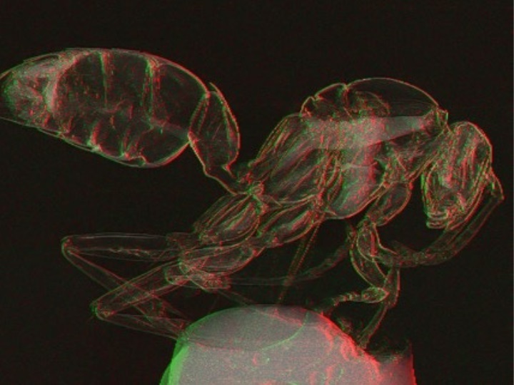
Groundbreaking X-Ray Imaging Technique Could Improve Medical Diagnostics
Posted on 19 Aug 2024
Older X-ray technology depends on the absorption of X-rays to generate images, which can be challenging when differentiating materials of similar density. This often results in low contrast images, making it difficult to distinguish between different substances, posing challenges in various applications including medical imaging. X-ray phase contrast imaging (PCI), which leverages relative phase changes as X-rays pass through an object, has become increasingly popular for its ability to enhance contrast, particularly for soft tissues. The single-mask differential method is notable among various PCI techniques for its simplicity and effectiveness, offering high-contrast imaging more efficiently and with lower radiation doses. Now, researchers have introduced a novel light transport model for a single-mask phase imaging system that enhances non-destructive deep imaging for visibility of light-element materials, including soft tissues such as cancers. This groundbreaking advancement in X-ray imaging technology could significantly improve medical diagnostics.
The new light transport model developed by researchers at the University of Houston (Houston, TX, USA) significantly enhances the capability of single-mask phase imaging systems, enabling non-destructive deep imaging that improves the visibility of light-element materials, such as soft tissues in medical diagnostics. This new model, detailed in a paper featured on the cover of Optica, facilitates the understanding of how contrast forms and how different contrast features interact within the acquired images. It enables the extraction of images that incorporate two distinct types of contrast mechanisms from a single exposure, marking a major improvement over conventional methods.

The innovative design utilizes an X-ray mask with periodic slits that align with the detector pixels to enhance edge contrast and capture differential phase information, clarifying variations between materials more distinctly. This advancement simplifies the imaging setup by reducing the reliance on high-resolution detectors or elaborate, multi-shot processes. The researchers have conducted extensive simulations and tested this model on a laboratory benchtop X-ray imaging system developed in-house. Their future objectives include adapting this technology for portable systems and retrofitting existing imaging setups, with plans to implement and evaluate it in practical settings such as hospitals.
“Our research opens up new possibilities for X-ray imaging by providing a simple, effective and low-cost method for enhancing image contrast which is a critical need for non-destructive deep imaging,” said Mini Das, Moores professor at UH’s College of Natural Sciences and Mathematics and Cullen College of Engineering. “It makes phase contrast imaging more accessible and practical, leading to better diagnostics and improved security screening. It is a versatile solution for a wide range of imaging challenges. We are in the process of testing the feasibility for a number of applications.”
Related Links:
University of Houston


