Optical Metabolic Imaging Identifies Breast Cancer Subtypes, Early Treatment Response
|
By MedImaging International staff writers Posted on 30 Oct 2013 |
Optical imaging technology, which measures metabolic activity in cancer cells, can accurately differentiate breast cancer subtypes, and it can detect responses to treatment as early as two days after therapy administration, according to recent research.
The study’s findings were published October 15, 2013, in Cancer Research, a journal of the American Association for Cancer Research. “The process of targeted drug development requires assays that measure drug target engagement and predict the response [or lack thereof] to treatment,” said Alex Walsh, a graduate student in the department of biomedical engineering at Vanderbilt University (Nashville, TN, USA). “We have shown that optical metabolic imaging [OMI] enables fast, sensitive, and accurate measurement of drug action. Importantly, OMI measurements can be made repeatedly over time in a live animal, which significantly reduces the cost of these preclinical studies.”
Human cells undergo extensive chemical reactions called metabolic activity to produce energy, and this activity is altered in cancer cells. Cancer cells’ metabolic activity changes when treated with anticancer drugs. OMI takes advantage of the fact that two molecules involved in cellular metabolism, called nicotinamide adenine dinucleotide (NADH) and flavin adenine dinucleotide (FAD), naturally emit fluorescence when exposed to specific forms of light. In this way, OMI generates distinctive signatures for cancer cells with a different metabolism and their responses to medications.
Dr. Walsh and coworkers used a custom-built, multiphoton microscope and coupled it with a titanium-sapphire laser that causes NADH and FAD to emit fluorescence. They employed specific filters to isolate the fluorescence emitted by these two molecules, and measured the ratio of the two as “redox ratio.” When they placed normal and cancerous breast cells under the microscope, OMI generated distinct signals for the two types of cells. OMI could also differentiate between estrogen receptor-positive, estrogen receptor-negative, HER2-positive, and HER2-negative breast cancer cells.
The researchers next examined the effect of the anti-HER2 antibody trastuzumab on three breast cancer-cell lines that respond differently to the antibody. They found that the redox ratios were significantly reduced in drug-sensitive cells after trastuzumab treatment but unaffected in the resistant cells. They then grew human breast tumors in mice and treated some of these with trastuzumab. When they imaged tumors in live mice, OMI demonstrated a difference in response between trastuzumab-sensitive and -resistant tumors as early as two days after the first dose of the antibody. Fluorodeoxyglucose-positron emission tomography (FDG-PET) imaging, in comparison, imaging, the conventional clinical metabolic imaging technique, could not measure any difference in response between trastuzumab-sensitive and -resistant tumors at any time point in the research, which lasted 12 days.
“Cancer drugs have profound effects on cellular energy production, and this can be harnessed by OMI to identify responding cells from nonresponding cells,” concluded Dr. Walsh. “We are hoping to develop a high-throughput screening method to predict the optimal drug treatment for a particular patient.”
Notably, OMI can be used on freshly excised tissues from patients; however, with additional refinements, it could be integrated in endoscopes for live imaging of human cancers, according to the investigators.
Related Links:
Vanderbilt University
Latest General/Advanced Imaging News
- CT Colonography Beats Stool DNA Testing for Colon Cancer Screening
- First-Of-Its-Kind Wearable Device Offers Revolutionary Alternative to CT Scans
- AI-Based CT Scan Analysis Predicts Early-Stage Kidney Damage Due to Cancer Treatments
- CT-Based Deep Learning-Driven Tool to Enhance Liver Cancer Diagnosis
- AI-Powered Imaging System Improves Lung Cancer Diagnosis
- AI Model Significantly Enhances Low-Dose CT Capabilities
- Ultra-Low Dose CT Aids Pneumonia Diagnosis in Immunocompromised Patients
- AI Reduces CT Lung Cancer Screening Workload by Almost 80%
- Cutting-Edge Technology Combines Light and Sound for Real-Time Stroke Monitoring
- AI System Detects Subtle Changes in Series of Medical Images Over Time
- New CT Scan Technique to Improve Prognosis and Treatments for Head and Neck Cancers
- World’s First Mobile Whole-Body CT Scanner to Provide Diagnostics at POC
- Comprehensive CT Scans Could Identify Atherosclerosis Among Lung Cancer Patients
- AI Improves Detection of Colorectal Cancer on Routine Abdominopelvic CT Scans
- Super-Resolution Technology Enhances Clinical Bone Imaging to Predict Osteoporotic Fracture Risk
- AI-Powered Abdomen Map Enables Early Cancer Detection
Channels
Radiography
view channel
RSNA AI Challenge Models Can Independently Interpret Mammograms
Breast cancer screening aims to detect cancers early while minimizing unnecessary recalls, yet balancing sensitivity and specificity remains a challenge. Automating detection in mammograms could help radiologists... Read more
New Technique Combines X-Ray Imaging and Radar for Safer Cancer Diagnosis
X-ray imaging methods, such as mammography and computed tomography (CT) scans, are essential for diagnosing and monitoring breast and lung cancer. While CT provides valuable three-dimensional insights,... Read moreMRI
view channel
AI-Assisted Model Enhances MRI Heart Scans
A cardiac MRI can reveal critical information about the heart’s function and any abnormalities, but traditional scans take 30 to 90 minutes and often suffer from poor image quality due to patient movement.... Read more
AI Model Outperforms Doctors at Identifying Patients Most At-Risk of Cardiac Arrest
Hypertrophic cardiomyopathy is one of the most common inherited heart conditions and a leading cause of sudden cardiac death in young individuals and athletes. While many patients live normal lives, some... Read moreUltrasound
view channel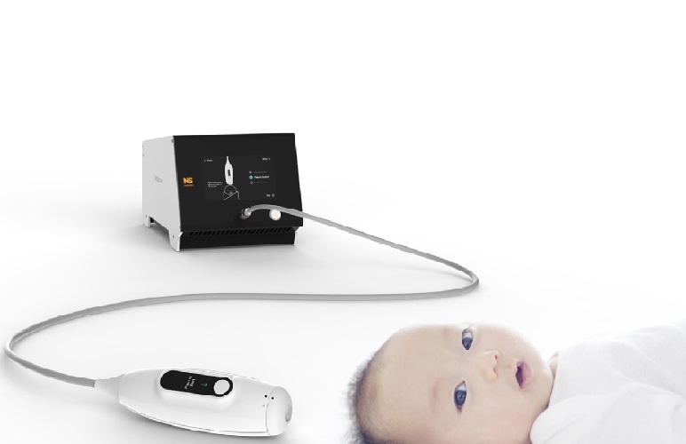
Non-Invasive Ultrasound-Based Tool Accurately Detects Infant Meningitis
Meningitis, an inflammation of the membranes surrounding the brain and spinal cord, can be fatal in infants if not diagnosed and treated early. Even when treated, it may leave lasting damage, such as cognitive... Read more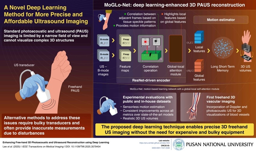
Breakthrough Deep Learning Model Enhances Handheld 3D Medical Imaging
Ultrasound imaging is a vital diagnostic technique used to visualize internal organs and tissues in real time and to guide procedures such as biopsies and injections. When paired with photoacoustic imaging... Read moreNuclear Medicine
view channel
Novel Bacteria-Specific PET Imaging Approach Detects Hard-To-Diagnose Lung Infections
Mycobacteroides abscessus is a rapidly growing mycobacteria that primarily affects immunocompromised patients and those with underlying lung diseases, such as cystic fibrosis or chronic obstructive pulmonary... Read more
New Imaging Approach Could Reduce Need for Biopsies to Monitor Prostate Cancer
Prostate cancer is the second leading cause of cancer-related death among men in the United States. However, the majority of older men diagnosed with prostate cancer have slow-growing, low-risk forms of... Read moreImaging IT
view channel
New Google Cloud Medical Imaging Suite Makes Imaging Healthcare Data More Accessible
Medical imaging is a critical tool used to diagnose patients, and there are billions of medical images scanned globally each year. Imaging data accounts for about 90% of all healthcare data1 and, until... Read more
Global AI in Medical Diagnostics Market to Be Driven by Demand for Image Recognition in Radiology
The global artificial intelligence (AI) in medical diagnostics market is expanding with early disease detection being one of its key applications and image recognition becoming a compelling consumer proposition... Read moreIndustry News
view channel
GE HealthCare and NVIDIA Collaboration to Reimagine Diagnostic Imaging
GE HealthCare (Chicago, IL, USA) has entered into a collaboration with NVIDIA (Santa Clara, CA, USA), expanding the existing relationship between the two companies to focus on pioneering innovation in... Read more
Patient-Specific 3D-Printed Phantoms Transform CT Imaging
New research has highlighted how anatomically precise, patient-specific 3D-printed phantoms are proving to be scalable, cost-effective, and efficient tools in the development of new CT scan algorithms... Read more
Siemens and Sectra Collaborate on Enhancing Radiology Workflows
Siemens Healthineers (Forchheim, Germany) and Sectra (Linköping, Sweden) have entered into a collaboration aimed at enhancing radiologists' diagnostic capabilities and, in turn, improving patient care... Read more


