SPECT/CT Technology Offers High Speed, Low Radiation Dose
|
By MedImaging International staff writers Posted on 28 Jun 2012 |

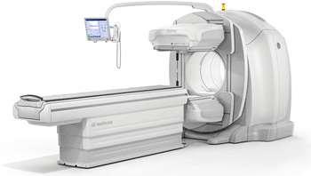
Image: The Optima NM/CT 640 system (Photo courtesy of GE Healthcare).
New imaging technology provides excellent single photon emission tomography/computed tomography (SPECT/CT) image quality enabled by high speed, one second CT rotation, minimizing motion artifacts while keeping radiation dose and ownership costs low.
At the Society of Nuclear Medicine held in Miami (FL, USA), in June 2012, GE Healthcare (Chalfont St. Giles, UK) introduced the latest addition to its Nuclear Medicine 600 series with a new performance SPECT/CT system, the Optima NM/CT 640, which offers nuclear medicine physicians the optimal balance of image quality, patient dose efficiency, and low total cost of ownership.
Based on the innovative 600 series SPECT technology found in the Discovery NM630, this system incorporates the latest generation general-purpose camera with a newly developed four-slice CT designed for hybrid instead of standalone CT use. The CT system, available in 2.5-mm and 5-mm slice thicknesses to optimize dose and resolution required for particular procedures, provides a typically low CT dose at 1-2 mSv for a 40-cm abdomen CT scan.
The Optima NM/CT 640 can be fully upgraded on location from a Discovery NM630 SPECT only system, and may be upgraded in the future to a 16-slice Discovery NM/CT 670, expanding not only its clinical capability, but offering the potential for research use. This upgradeability helps protect clinicians’ and healthcare providers’ investments as the needs of their department evolve.
With its small footprint (5.7 m x 3.6 m) the Optima NM/CT 640 requires minimal renovation and installation costs. With the benefit of optimized CT power, shielding, and control room requirements are often eliminated; saving as much as USD 100,000 compared to higher CT powered systems.
“At GE Healthcare, we are dedicated to pushing nuclear medicine to its full potential and investing in its future,” said Nathan Hermony, general manager, nuclear medicine, GE Healthcare. “We’re focused on developing equipment that helps customers address the challenges they are confronted with every day--high image quality, low dose, and short exam times. Adding the Optima NM/CT 640 to our portfolio helps us strengthen this position, allows customers to upgrade as their needs expand, and continues to benefit clinicians and their patients.”
Adding all the benefits of the Xeleris Workstation with the Evolution technology to the Optima NM/CT 640 reduces the trade-offs that are frequently required between acquisition time, dose and image quality. By allowing clinicians to feel confident in their diagnosis, when reducing time or injected patient dose by up to 50% in most scanning procedures while still maintaining excellent image quality.
The Xeleris workstation--which integrates new and existing nuclear medicine equipment, including legacy GE and non-GE devices--is designed to provide consistent results and enhanced workflow. Xeleris can keep clinicians connected to images and applications from picture archiving and communication systems (PACS) and personal computers (PCs) with their institution and remotely.
The Optima NM/CT 640 is engineered to accommodate more patients than previous generation nuclear medicine systems. With its 70-cm-wide bore and table capable of handling patients up to 227 kg, the Optima NM/CT 640 provides access to a wide variety of patients.
“Building off of our extensive experience in SPECT/CT, the advances we’ve made to our Infinia Hawkeye 4 platform and incorporating the SPECT technology of our Discovery NM630 camera, we’re striving to give our customers unsurpassed diagnostic confidence,” added Mr. Hermony.
Related Links:
GE Healthcare
Latest Nuclear Medicine News
- Novel Radiolabeled Antibody Improves Diagnosis and Treatment of Solid Tumors
- Novel PET Imaging Approach Offers Never-Before-Seen View of Neuroinflammation
- Novel Radiotracer Identifies Biomarker for Triple-Negative Breast Cancer
- Innovative PET Imaging Technique to Help Diagnose Neurodegeneration
- New Molecular Imaging Test to Improve Lung Cancer Diagnosis
- Novel PET Technique Visualizes Spinal Cord Injuries to Predict Recovery
- Next-Gen Tau Radiotracers Outperform FDA-Approved Imaging Agents in Detecting Alzheimer’s
- Breakthrough Method Detects Inflammation in Body Using PET Imaging
- Advanced Imaging Reveals Hidden Metastases in High-Risk Prostate Cancer Patients
- Combining Advanced Imaging Technologies Offers Breakthrough in Glioblastoma Treatment
- New Molecular Imaging Agent Accurately Identifies Crucial Cancer Biomarker
- New Scans Light Up Aggressive Tumors for Better Treatment
- AI Stroke Brain Scan Readings Twice as Accurate as Current Method
- AI Analysis of PET/CT Images Predicts Side Effects of Immunotherapy in Lung Cancer
- New Imaging Agent to Drive Step-Change for Brain Cancer Imaging
- Portable PET Scanner to Detect Earliest Stages of Alzheimer’s Disease
Channels
Radiography
view channel
World's Largest Class Single Crystal Diamond Radiation Detector Opens New Possibilities for Diagnostic Imaging
Diamonds possess ideal physical properties for radiation detection, such as exceptional thermal and chemical stability along with a quick response time. Made of carbon with an atomic number of six, diamonds... Read more
AI-Powered Imaging Technique Shows Promise in Evaluating Patients for PCI
Percutaneous coronary intervention (PCI), also known as coronary angioplasty, is a minimally invasive procedure where small metal tubes called stents are inserted into partially blocked coronary arteries... Read moreMRI
view channel
AI Tool Predicts Relapse of Pediatric Brain Cancer from Brain MRI Scans
Many pediatric gliomas are treatable with surgery alone, but relapses can be catastrophic. Predicting which patients are at risk for recurrence remains challenging, leading to frequent follow-ups with... Read more
AI Tool Tracks Effectiveness of Multiple Sclerosis Treatments Using Brain MRI Scans
Multiple sclerosis (MS) is a condition in which the immune system attacks the brain and spinal cord, leading to impairments in movement, sensation, and cognition. Magnetic Resonance Imaging (MRI) markers... Read more
Ultra-Powerful MRI Scans Enable Life-Changing Surgery in Treatment-Resistant Epileptic Patients
Approximately 360,000 individuals in the UK suffer from focal epilepsy, a condition in which seizures spread from one part of the brain. Around a third of these patients experience persistent seizures... Read moreUltrasound
view channel.jpeg)
AI-Powered Lung Ultrasound Outperforms Human Experts in Tuberculosis Diagnosis
Despite global declines in tuberculosis (TB) rates in previous years, the incidence of TB rose by 4.6% from 2020 to 2023. Early screening and rapid diagnosis are essential elements of the World Health... Read more
AI Identifies Heart Valve Disease from Common Imaging Test
Tricuspid regurgitation is a condition where the heart's tricuspid valve does not close completely during contraction, leading to backward blood flow, which can result in heart failure. A new artificial... Read moreGeneral/Advanced Imaging
view channel
AI-Powered Imaging System Improves Lung Cancer Diagnosis
Given the need to detect lung cancer at earlier stages, there is an increasing need for a definitive diagnostic pathway for patients with suspicious pulmonary nodules. However, obtaining tissue samples... Read more
AI Model Significantly Enhances Low-Dose CT Capabilities
Lung cancer remains one of the most challenging diseases, making early diagnosis vital for effective treatment. Fortunately, advancements in artificial intelligence (AI) are revolutionizing lung cancer... Read moreImaging IT
view channel
New Google Cloud Medical Imaging Suite Makes Imaging Healthcare Data More Accessible
Medical imaging is a critical tool used to diagnose patients, and there are billions of medical images scanned globally each year. Imaging data accounts for about 90% of all healthcare data1 and, until... Read more
Global AI in Medical Diagnostics Market to Be Driven by Demand for Image Recognition in Radiology
The global artificial intelligence (AI) in medical diagnostics market is expanding with early disease detection being one of its key applications and image recognition becoming a compelling consumer proposition... Read moreIndustry News
view channel
GE HealthCare and NVIDIA Collaboration to Reimagine Diagnostic Imaging
GE HealthCare (Chicago, IL, USA) has entered into a collaboration with NVIDIA (Santa Clara, CA, USA), expanding the existing relationship between the two companies to focus on pioneering innovation in... Read more
Patient-Specific 3D-Printed Phantoms Transform CT Imaging
New research has highlighted how anatomically precise, patient-specific 3D-printed phantoms are proving to be scalable, cost-effective, and efficient tools in the development of new CT scan algorithms... Read more
Siemens and Sectra Collaborate on Enhancing Radiology Workflows
Siemens Healthineers (Forchheim, Germany) and Sectra (Linköping, Sweden) have entered into a collaboration aimed at enhancing radiologists' diagnostic capabilities and, in turn, improving patient care... Read more


