Better Designed Ultrasound Could Improve Image Clarity
|
By MedImaging International staff writers Posted on 26 Jun 2012 |

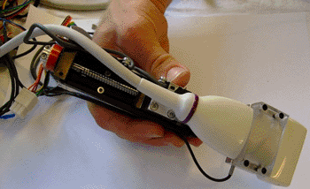
Image: The handheld, force-controlled ultrasound (Photo courtesy of MIT).
New ultrasound technology allows precise measurements and tracking of disease progression.
New research conducted at the Massachusetts Institute of Technology (MIT; Cambridge, MA, USA) could improve the ability of untrained workers to perform fundamental ultrasound scanning, while allowing trained workers to much more effectively monitor the development of medical conditions, such as the accumulation of plaque in arteries or the growth of a tumor.
The improvements to this widely used technology could provide detailed information far beyond what is possible with existing systems, according to the researchers. The research, led by Dr. Brian W. Anthony, codirector of MIT’s Medical Electronic Device Realization Center (MEDRC) and director of the Master of Engineering in Manufacturing Program, was presented in May 2012 at the International Symposium on Biomedical Imaging in Barcelona (Spain).
There are two major features to the improvements engineered by Dr. Anthony and his team. First, the researchers developed a way to adjust for variations in the force exerted by a sonographer, generating more consistent images that can compensate for body motions such as breathing and heartbeat. Second, they provided a way to map the precise location on the skin where one reading was taken, so that it can be precisely matched with later readings to identify changes in the size or location of a tumor, clot, or other structure.
Together, the two improvements could make ultrasound a much more precise application for monitoring the progression of disease, according to Dr. Anthony. The devices are currently undergoing three clinical trials, including one at Boston Children’s Hospital (MA, USA) focused on monitoring the progression of patients with Duchenne muscular dystrophy (DMD).
In that trial, Dr. Anthony reported, researchers are trying to determine “how fast the muscle deteriorates, and how effective different medications are.” It’s important to have a reliable way of monitoring changes in muscle. The study is aimed at determining whether ultrasound imaging can serve as a convenient, noninvasive, clinically meaningful way of monitoring disease progression in DMD.
The new device maintains constant force through the addition of a force sensor to its probe tip and servomotors that can respond almost instantly to changes in force. That, in turn, makes it possible to examine how the image varies as the force increases, which can provide important diagnostic information about the elasticity of skin, muscle, and other tissues.
To provide accurate positioning, a tiny camera and lens mounted on the probe can reveal skin patterns that are distinctive and constant, similar to fingerprints. “Skin patterns are pretty unique,” Dr. Anthony said, the system, utilizing software to compare new images with earlier ones, “can get you back to that same patch of skin,” something that is impossible to do manually.
Dr. Anthony compares that precise positioning to “an on-the-patient global positioning system (GPS) system” for locating structures in the body. The ability to take images over time from exactly the same position makes it possible to monitor changing tissues quite precisely: The imaging system can determine the volume of a near-surface tumor or other feature to within an accuracy of 1%-2%, according to Dr. Anthony. There are existing ways to get this kind of accuracy, but these require expensive specialized equipment that few hospitals have.
Besides the potential for these sophisticated diagnostic capabilities, enhanced control over testing could make it possible for relatively untrained health care workers to administer basic ultrasound pregnancy tests--particularly in remote, underserved areas where trained sonographers may not be available. The various control methods “take the uncertainty out” of the process, Anthony says.
Dr. Craig Steiner, an anesthesiologist at Chester County Hospital (PA, USA), said, “I’m excited about the prospects” of these improved systems. “The reproducibility of the scan with consistent pressure and picture quality would help with remote readings of locally done scans. This could be relevant for teleradiology, which is an area ripe for expansion. The field of ultrasound is still developing. Ultrasound will partially replace CT scans, reduce radiation exposure to patients, and make diagnosing easier when away from the high-cost hospitals. It can help our world provide care at a more reasonable cost with a new paradigm of care.”
Related Links:
Massachusetts Institute of Technology
Latest Ultrasound News
- AI-Powered Lung Ultrasound Outperforms Human Experts in Tuberculosis Diagnosis
- AI Identifies Heart Valve Disease from Common Imaging Test
- Novel Imaging Method Enables Early Diagnosis and Treatment Monitoring of Type 2 Diabetes
- Ultrasound-Based Microscopy Technique to Help Diagnose Small Vessel Diseases
- Smart Ultrasound-Activated Immune Cells Destroy Cancer Cells for Extended Periods
- Tiny Magnetic Robot Takes 3D Scans from Deep Within Body
- High Resolution Ultrasound Speeds Up Prostate Cancer Diagnosis
- World's First Wireless, Handheld, Whole-Body Ultrasound with Single PZT Transducer Makes Imaging More Accessible
- Artificial Intelligence Detects Undiagnosed Liver Disease from Echocardiograms
- Ultrasound Imaging Non-Invasively Tracks Tumor Response to Radiation and Immunotherapy
- AI Improves Detection of Congenital Heart Defects on Routine Prenatal Ultrasounds
- AI Diagnoses Lung Diseases from Ultrasound Videos with 96.57% Accuracy
- New Contrast Agent for Ultrasound Imaging Ensures Affordable and Safer Medical Diagnostics
- Ultrasound-Directed Microbubbles Boost Immune Response Against Tumors
- POC Ultrasound Enhances Early Pregnancy Care and Cuts Emergency Visits
- AI-Based Models Outperform Human Experts at Identifying Ovarian Cancer in Ultrasound Images
Channels
Radiography
view channel
AI Improves Early Detection of Interval Breast Cancers
Interval breast cancers, which occur between routine screenings, are easier to treat when detected earlier. Early detection can reduce the need for aggressive treatments and improve the chances of better outcomes.... Read more
World's Largest Class Single Crystal Diamond Radiation Detector Opens New Possibilities for Diagnostic Imaging
Diamonds possess ideal physical properties for radiation detection, such as exceptional thermal and chemical stability along with a quick response time. Made of carbon with an atomic number of six, diamonds... Read moreMRI
view channel
New MRI Technique Reveals True Heart Age to Prevent Attacks and Strokes
Heart disease remains one of the leading causes of death worldwide. Individuals with conditions such as diabetes or obesity often experience accelerated aging of their hearts, sometimes by decades.... Read more
AI Tool Predicts Relapse of Pediatric Brain Cancer from Brain MRI Scans
Many pediatric gliomas are treatable with surgery alone, but relapses can be catastrophic. Predicting which patients are at risk for recurrence remains challenging, leading to frequent follow-ups with... Read more
AI Tool Tracks Effectiveness of Multiple Sclerosis Treatments Using Brain MRI Scans
Multiple sclerosis (MS) is a condition in which the immune system attacks the brain and spinal cord, leading to impairments in movement, sensation, and cognition. Magnetic Resonance Imaging (MRI) markers... Read more
Ultra-Powerful MRI Scans Enable Life-Changing Surgery in Treatment-Resistant Epileptic Patients
Approximately 360,000 individuals in the UK suffer from focal epilepsy, a condition in which seizures spread from one part of the brain. Around a third of these patients experience persistent seizures... Read moreNuclear Medicine
view channel
Novel Radiolabeled Antibody Improves Diagnosis and Treatment of Solid Tumors
Interleukin-13 receptor α-2 (IL13Rα2) is a cell surface receptor commonly found in solid tumors such as glioblastoma, melanoma, and breast cancer. It is minimally expressed in normal tissues, making it... Read more
Novel PET Imaging Approach Offers Never-Before-Seen View of Neuroinflammation
COX-2, an enzyme that plays a key role in brain inflammation, can be significantly upregulated by inflammatory stimuli and neuroexcitation. Researchers suggest that COX-2 density in the brain could serve... Read moreGeneral/Advanced Imaging
view channel
CT-Based Deep Learning-Driven Tool to Enhance Liver Cancer Diagnosis
Medical imaging, such as computed tomography (CT) scans, plays a crucial role in oncology, offering essential data for cancer detection, treatment planning, and monitoring of response to therapies.... Read more
AI-Powered Imaging System Improves Lung Cancer Diagnosis
Given the need to detect lung cancer at earlier stages, there is an increasing need for a definitive diagnostic pathway for patients with suspicious pulmonary nodules. However, obtaining tissue samples... Read moreImaging IT
view channel
New Google Cloud Medical Imaging Suite Makes Imaging Healthcare Data More Accessible
Medical imaging is a critical tool used to diagnose patients, and there are billions of medical images scanned globally each year. Imaging data accounts for about 90% of all healthcare data1 and, until... Read more
Global AI in Medical Diagnostics Market to Be Driven by Demand for Image Recognition in Radiology
The global artificial intelligence (AI) in medical diagnostics market is expanding with early disease detection being one of its key applications and image recognition becoming a compelling consumer proposition... Read moreIndustry News
view channel
GE HealthCare and NVIDIA Collaboration to Reimagine Diagnostic Imaging
GE HealthCare (Chicago, IL, USA) has entered into a collaboration with NVIDIA (Santa Clara, CA, USA), expanding the existing relationship between the two companies to focus on pioneering innovation in... Read more
Patient-Specific 3D-Printed Phantoms Transform CT Imaging
New research has highlighted how anatomically precise, patient-specific 3D-printed phantoms are proving to be scalable, cost-effective, and efficient tools in the development of new CT scan algorithms... Read more
Siemens and Sectra Collaborate on Enhancing Radiology Workflows
Siemens Healthineers (Forchheim, Germany) and Sectra (Linköping, Sweden) have entered into a collaboration aimed at enhancing radiologists' diagnostic capabilities and, in turn, improving patient care... Read more


