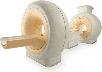
Fusion Imaging System Designed for Standalone MR and Hybrid PET/MR Imaging
|
By MedImaging International staff writers Posted on 27 Dec 2011 |

Image: Ingenuity TF PET/MR (Photo courtesy of Philips Healthcare).
New whole body positron emission tomography/magnetic resonance (PET/MR) technology provides increased economic value because it is a sequential imaging system that has a similar clinical workflow experience to PET/computed tomography (CT), the current standard for hybrid imaging.
Moreover, the system is designed so the patient table rotates between each modality to scan a patient, thereby enabling the system to perform both standalone MR and hybrid PET/MR studies. This provides added flexibility by eliminating the need to invest in multiple scanners while decreasing throughput time and improving patient comfort since the patient can remain on the same table for both tests.
Philips Healthcare (Best, The Netherlands) announced 510(k) clearance from the US Food and Drug Administration (FDA) for the company’s first commercially available whole body PET/MR imaging system, the Ingenuity TF PET/MR. This innovative platform should revolutionize how medicine is practiced in the future by helping clinicians and researchers study novel customized medicine and treatments for oncology, cardiology, and neurology. The system was presented at the 97th annual meeting of the Radiological Society of North America (RSNA), November 27-December 2, 2010.
It was previously thought that PET and MR scans were incompatible; however, Philips overcame the enormous technical obstacles, through advances in technology, to create a new class of hybrid imaging that should push the boundary of what is possible in imaging. The system is designed to provide a cutting-edge platform well into the future by simplifying the addition of new technologies as they become available.
Studies on the Ingenuity TF PET/MR have shown that bringing high fidelity PET and MR imaging together provides high quality diagnostic images. “The PET/MR system will be useful to practitioners because of the highly anatomical and contrast images that can be acquired when you combine functional MR images with the metabolic information acquired by PET,” said Zahi Fayad, PhD, professor of radiology and medicine (cardiology), and director, Translational and Molecular Imaging Institute, at the Mount Sinai School of Medicine (New York, NY, USA).
Clinical cases have already shown the benefits of being able to superimpose PET over MR images to help detect abnormalities in various organs. Previously, this was not possible because the two studies took place at different times, with different conditions and with different patient positions. In addition to the possibility of expanding clinical prospects as an advanced research tool, the technology could also be used in a clinical setting to support a patient’s entire care cycle process from detection or diagnosis to long-term disease management.
The system features Philips’ proprietary time-of-flight (TOF) technology, Astonish TF, a technology for PET scanners that is designed to enhance image quality by reducing noise and providing increased sensitivity. It is combined with an excellent soft-tissue contrast of 3T MR to image disease as it proliferates in soft tissue.
“Over the years, the introduction of new medical imaging technologies has helped to expand clinical horizons,” said Gene Saragnese, executive vice-president and CEO, imaging systems, for Philips Healthcare. “The Ingenuity TF PET/MR is a state-of-the-art platform that will remain state-of-the-art as it continues to evolve over time to provide robust research and clinical value. This will change the way health care is practiced in the future.”
Mount Sinai and University Hospitals/Case Western Reserve University (Cleveland, OH, USA) will house the first Philips combined, whole-body PET/MR systems in the United States. “We are specifically interested in PET/MR because the combination is expected to provide a more advanced understanding of the processes taking place in vascular beds,” said Dr. Fayad.
With the program formally launched in 2007, Philips’ PET/MR system is the embodiment of hybrid imaging, a major trend that is a growth driver of imaging procedure volumes. Philips was the first company to bring a PET/MR system to market when CE Mark in Europe was earned in January 2011. With FDA clearance, the system can now be marketed in the world’s largest healthcare market.
Related Links:
Philips Healthcare
Latest Nuclear Medicine News
- Novel Radiolabeled Antibody Improves Diagnosis and Treatment of Solid Tumors
- Novel PET Imaging Approach Offers Never-Before-Seen View of Neuroinflammation
- Novel Radiotracer Identifies Biomarker for Triple-Negative Breast Cancer
- Innovative PET Imaging Technique to Help Diagnose Neurodegeneration
- New Molecular Imaging Test to Improve Lung Cancer Diagnosis
- Novel PET Technique Visualizes Spinal Cord Injuries to Predict Recovery
- Next-Gen Tau Radiotracers Outperform FDA-Approved Imaging Agents in Detecting Alzheimer’s
- Breakthrough Method Detects Inflammation in Body Using PET Imaging
- Advanced Imaging Reveals Hidden Metastases in High-Risk Prostate Cancer Patients
- Combining Advanced Imaging Technologies Offers Breakthrough in Glioblastoma Treatment
- New Molecular Imaging Agent Accurately Identifies Crucial Cancer Biomarker
- New Scans Light Up Aggressive Tumors for Better Treatment
- AI Stroke Brain Scan Readings Twice as Accurate as Current Method
- AI Analysis of PET/CT Images Predicts Side Effects of Immunotherapy in Lung Cancer
- New Imaging Agent to Drive Step-Change for Brain Cancer Imaging
- Portable PET Scanner to Detect Earliest Stages of Alzheimer’s Disease
Channels
Radiography
view channel
AI Improves Early Detection of Interval Breast Cancers
Interval breast cancers, which occur between routine screenings, are easier to treat when detected earlier. Early detection can reduce the need for aggressive treatments and improve the chances of better outcomes.... Read more
World's Largest Class Single Crystal Diamond Radiation Detector Opens New Possibilities for Diagnostic Imaging
Diamonds possess ideal physical properties for radiation detection, such as exceptional thermal and chemical stability along with a quick response time. Made of carbon with an atomic number of six, diamonds... Read moreMRI
view channel
Cutting-Edge MRI Technology to Revolutionize Diagnosis of Common Heart Problem
Aortic stenosis is a common and potentially life-threatening heart condition. It occurs when the aortic valve, which regulates blood flow from the heart to the rest of the body, becomes stiff and narrow.... Read more
New MRI Technique Reveals True Heart Age to Prevent Attacks and Strokes
Heart disease remains one of the leading causes of death worldwide. Individuals with conditions such as diabetes or obesity often experience accelerated aging of their hearts, sometimes by decades.... Read more
AI Tool Predicts Relapse of Pediatric Brain Cancer from Brain MRI Scans
Many pediatric gliomas are treatable with surgery alone, but relapses can be catastrophic. Predicting which patients are at risk for recurrence remains challenging, leading to frequent follow-ups with... Read more
AI Tool Tracks Effectiveness of Multiple Sclerosis Treatments Using Brain MRI Scans
Multiple sclerosis (MS) is a condition in which the immune system attacks the brain and spinal cord, leading to impairments in movement, sensation, and cognition. Magnetic Resonance Imaging (MRI) markers... Read moreUltrasound
view channel.jpeg)
AI-Powered Lung Ultrasound Outperforms Human Experts in Tuberculosis Diagnosis
Despite global declines in tuberculosis (TB) rates in previous years, the incidence of TB rose by 4.6% from 2020 to 2023. Early screening and rapid diagnosis are essential elements of the World Health... Read more
AI Identifies Heart Valve Disease from Common Imaging Test
Tricuspid regurgitation is a condition where the heart's tricuspid valve does not close completely during contraction, leading to backward blood flow, which can result in heart failure. A new artificial... Read moreGeneral/Advanced Imaging
view channel
AI-Based CT Scan Analysis Predicts Early-Stage Kidney Damage Due to Cancer Treatments
Radioligand therapy, a form of targeted nuclear medicine, has recently gained attention for its potential in treating specific types of tumors. However, one of the potential side effects of this therapy... Read more
CT-Based Deep Learning-Driven Tool to Enhance Liver Cancer Diagnosis
Medical imaging, such as computed tomography (CT) scans, plays a crucial role in oncology, offering essential data for cancer detection, treatment planning, and monitoring of response to therapies.... Read moreImaging IT
view channel
New Google Cloud Medical Imaging Suite Makes Imaging Healthcare Data More Accessible
Medical imaging is a critical tool used to diagnose patients, and there are billions of medical images scanned globally each year. Imaging data accounts for about 90% of all healthcare data1 and, until... Read more
Global AI in Medical Diagnostics Market to Be Driven by Demand for Image Recognition in Radiology
The global artificial intelligence (AI) in medical diagnostics market is expanding with early disease detection being one of its key applications and image recognition becoming a compelling consumer proposition... Read moreIndustry News
view channel
GE HealthCare and NVIDIA Collaboration to Reimagine Diagnostic Imaging
GE HealthCare (Chicago, IL, USA) has entered into a collaboration with NVIDIA (Santa Clara, CA, USA), expanding the existing relationship between the two companies to focus on pioneering innovation in... Read more
Patient-Specific 3D-Printed Phantoms Transform CT Imaging
New research has highlighted how anatomically precise, patient-specific 3D-printed phantoms are proving to be scalable, cost-effective, and efficient tools in the development of new CT scan algorithms... Read more
Siemens and Sectra Collaborate on Enhancing Radiology Workflows
Siemens Healthineers (Forchheim, Germany) and Sectra (Linköping, Sweden) have entered into a collaboration aimed at enhancing radiologists' diagnostic capabilities and, in turn, improving patient care... Read more


 Guided Devices.jpg)


