4D Cardiovascular Ultrasound Technology Improves Image Quality, Quantification, and Productivity
|
By MedImaging International staff writers Posted on 22 Nov 2010 |

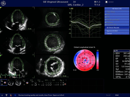
Image: A dilated heart shown with 4D strain (photo courtesy Business Wire).
A new development--for the first time in cardiovascular ultrasound--provides dynamic multislice acquisition technology with the ability to simultaneously acquire images of up to 12 slices of the heart, and automated function imaging (AFI) for tri-plane measurements. The technology also includes new quantification tools including four-dimensional (4D) strain, 2D auto ejection fraction (EF), and AFI for transeshophageal echocardiography (TEE), for left ventricular (LV), global, and regional function.
GE Healthcare (Chalfont St. Giles, UK), a unit of General Electric Company (Fairfield, CT, USA), has received U.S. Food and Drug Administration (FDA) 510(k) clearance for the latest version of its built-for-4D Vivid E9 cardiovascular ultrasound system. Vivid E9 Breakthrough 2011 (BT11) provides innovative features designed to help improve image quality, quantification, and workflow.
Vivid E9 BT11 is now FDA-cleared, CE Marked, and available for sale in many Asian and South American countries. The technology is available in 2D or 4D configuration with support for a comprehensive range of adult and pediatric probes. New features improve image quality, quantification, and workflow when compared to the previous generation of Vivid E9 and include: a new high bandwidth 4V-D transducer, significantly smaller than its 3V-D predecessor, yet more powerful, with improved 2D and 4D image quality and the ability to scan in all modes, including continuous wave (CW) Doppler; a new 12S-D transducer for the smallest of neonatal patients, helping enhance a range of pediatric echo transducers available for Vivid E9; improved performance and expanded application coverage for shared services with the addition of a ML6-15-D probe, and in the operating room with the intraoperative i13L probe.
Other system features include a new and improved 4D Auto LVQ, an integrated and automated 4D volume and EF tool, and, an extension to this tool, 4D LV Mass, which provides a left ventricular mass, as well as a mass index; 4D Strain, an easy-to-interpret 4D tracking tool that allows for quantification of left ventricular function, providing global and regional longitudinal, circumferential, radial, and area strain curves as well as bulls-eyes to possibly identify wall motion abnormalities.
Scan Assist and Scan Assist Pro, with its customizable protocol driven exam, aids users in acquiring the required images in a predefined sequence and can considerably reduce keystrokes and exam time, providing opportunities for improved workflow and potential productivity gains. Multidimensional imaging allows users to acquire bi- or tri-plane images in a single beat, which helps reduce acquisition time and storage space.
The system's Auto-Align for 4D views features fully automatic positioning and orientation of the left ventricle, helping to improve efficiency and consistency of image analysis. Multislice imaging provides a live imaging mode with choices of viewing 5, 7, 9, or 12 slices for simultaneous acquisition and assessment. Dynamic multislice provides for continuous visualization of the same cardiac structures throughout the heart cycle, potentially improving the accuracy of wall motion assessment of multislice images.
Related Links:
GE Healthcare
Latest Ultrasound News
- New Incision-Free Technique Halts Growth of Debilitating Brain Lesions
- AI-Powered Lung Ultrasound Outperforms Human Experts in Tuberculosis Diagnosis
- AI Identifies Heart Valve Disease from Common Imaging Test
- Novel Imaging Method Enables Early Diagnosis and Treatment Monitoring of Type 2 Diabetes
- Ultrasound-Based Microscopy Technique to Help Diagnose Small Vessel Diseases
- Smart Ultrasound-Activated Immune Cells Destroy Cancer Cells for Extended Periods
- Tiny Magnetic Robot Takes 3D Scans from Deep Within Body
- High Resolution Ultrasound Speeds Up Prostate Cancer Diagnosis
- World's First Wireless, Handheld, Whole-Body Ultrasound with Single PZT Transducer Makes Imaging More Accessible
- Artificial Intelligence Detects Undiagnosed Liver Disease from Echocardiograms
- Ultrasound Imaging Non-Invasively Tracks Tumor Response to Radiation and Immunotherapy
- AI Improves Detection of Congenital Heart Defects on Routine Prenatal Ultrasounds
- AI Diagnoses Lung Diseases from Ultrasound Videos with 96.57% Accuracy
- New Contrast Agent for Ultrasound Imaging Ensures Affordable and Safer Medical Diagnostics
- Ultrasound-Directed Microbubbles Boost Immune Response Against Tumors
- POC Ultrasound Enhances Early Pregnancy Care and Cuts Emergency Visits
Channels
Radiography
view channel
Machine Learning Algorithm Identifies Cardiovascular Risk from Routine Bone Density Scans
A new study published in the Journal of Bone and Mineral Research reveals that an automated machine learning program can predict the risk of cardiovascular events and falls or fractures by analyzing bone... Read more
AI Improves Early Detection of Interval Breast Cancers
Interval breast cancers, which occur between routine screenings, are easier to treat when detected earlier. Early detection can reduce the need for aggressive treatments and improve the chances of better outcomes.... Read more
World's Largest Class Single Crystal Diamond Radiation Detector Opens New Possibilities for Diagnostic Imaging
Diamonds possess ideal physical properties for radiation detection, such as exceptional thermal and chemical stability along with a quick response time. Made of carbon with an atomic number of six, diamonds... Read moreMRI
view channel
New MRI Technique Reveals Hidden Heart Issues
Traditional exercise stress tests conducted within an MRI machine require patients to lie flat, a position that artificially improves heart function by increasing stroke volume due to gravity-driven blood... Read more
Shorter MRI Exam Effectively Detects Cancer in Dense Breasts
Women with extremely dense breasts face a higher risk of missed breast cancer diagnoses, as dense glandular and fibrous tissue can obscure tumors on mammograms. While breast MRI is recommended for supplemental... Read moreNuclear Medicine
view channel
New Imaging Approach Could Reduce Need for Biopsies to Monitor Prostate Cancer
Prostate cancer is the second leading cause of cancer-related death among men in the United States. However, the majority of older men diagnosed with prostate cancer have slow-growing, low-risk forms of... Read more
Novel Radiolabeled Antibody Improves Diagnosis and Treatment of Solid Tumors
Interleukin-13 receptor α-2 (IL13Rα2) is a cell surface receptor commonly found in solid tumors such as glioblastoma, melanoma, and breast cancer. It is minimally expressed in normal tissues, making it... Read moreGeneral/Advanced Imaging
view channel
First-Of-Its-Kind Wearable Device Offers Revolutionary Alternative to CT Scans
Currently, patients with conditions such as heart failure, pneumonia, or respiratory distress often require multiple imaging procedures that are intermittent, disruptive, and involve high levels of radiation.... Read more
AI-Based CT Scan Analysis Predicts Early-Stage Kidney Damage Due to Cancer Treatments
Radioligand therapy, a form of targeted nuclear medicine, has recently gained attention for its potential in treating specific types of tumors. However, one of the potential side effects of this therapy... Read moreImaging IT
view channel
New Google Cloud Medical Imaging Suite Makes Imaging Healthcare Data More Accessible
Medical imaging is a critical tool used to diagnose patients, and there are billions of medical images scanned globally each year. Imaging data accounts for about 90% of all healthcare data1 and, until... Read more
Global AI in Medical Diagnostics Market to Be Driven by Demand for Image Recognition in Radiology
The global artificial intelligence (AI) in medical diagnostics market is expanding with early disease detection being one of its key applications and image recognition becoming a compelling consumer proposition... Read moreIndustry News
view channel
GE HealthCare and NVIDIA Collaboration to Reimagine Diagnostic Imaging
GE HealthCare (Chicago, IL, USA) has entered into a collaboration with NVIDIA (Santa Clara, CA, USA), expanding the existing relationship between the two companies to focus on pioneering innovation in... Read more
Patient-Specific 3D-Printed Phantoms Transform CT Imaging
New research has highlighted how anatomically precise, patient-specific 3D-printed phantoms are proving to be scalable, cost-effective, and efficient tools in the development of new CT scan algorithms... Read more
Siemens and Sectra Collaborate on Enhancing Radiology Workflows
Siemens Healthineers (Forchheim, Germany) and Sectra (Linköping, Sweden) have entered into a collaboration aimed at enhancing radiologists' diagnostic capabilities and, in turn, improving patient care... Read more


