PET Technology Devised for Heart Imaging
|
By MedImaging International staff writers Posted on 12 Apr 2010 |

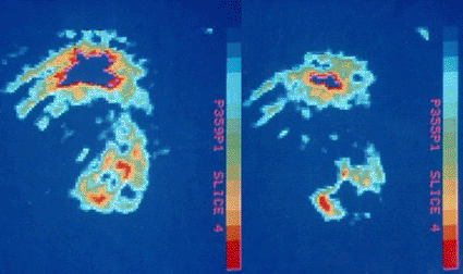
Image: PET scan of the human heart showing the effect of a blood clot (Photo courtesy of Science Source).
A new positron emission tomography (PET) system was developed and optimized for molecular imaging of the heart, making it a suitable solution for cardiologists and hospitals looking to add high accuracy, cost effective imaging technology.
The nuclear cardiology imaging scene has been dominated by single photon emission tomography (SPECT) until recently when the U.S. imaging world was shaken by the announcement of SPECT reimbursement cuts by the U.S. Centers for Medicare and Medicaid Services (CMS), combined with the world shortage of the molybdenum-99 isotope. Many in the industry are looking for new technologies to improve their diagnostic accuracy, improve patient outcomes, reduce patient radiation exposure all while adding to their bottom line. The elusive solution to this dismal situation may lie in an already well-established underutilized imaging modality: PET.
While PET is a more costly procedure than the SPECT imaging, the use of PET in cardiac nuclear medicine has been shown to reduce long-term costs and resolve clinically complicated cases. The accuracy of PET helps reduce the need for unnecessary angiograms. It can also reduce bypass surgeries by more accurately risk-stratifying patients that may require the invasive procedure from those that might benefit from alternative therapies. This modality has also been shown to monitor therapy quantitatively, which helps provide personalized medicine plan for each patient. PET, specifically without a computed tomography (CT), has shown to have the lowest radiation exposure for the assessment of coronary disease.
Taking advantage of these trends, Positron Corp. (Fishers, IN, USA) strategically introduced the industry's first cardiac optimized PET scanner, Attrius. The scanner is designed to provide a significantly lower cost of ownership as compared to PET/CT modalities and does not need additional space for electronics. It has a much smaller footprint, fewer boards, easier access to the detector modules, less power consumption, and automated tuning features. The product can easily integrate into practices of all sizes. The table limit was increased to 204 kg, permitting larger patients to be imaged. The table is also capable of loading patients from the front or back, improving the position options for imaging.
Furthermore, Positron's cardiac PET scanner is one of the highest two-dimensional (2D) sensitivity systems on the market today. It features more uniformity achieved in its slice sensitivity, consistency in the quantitation from slice-to-slice, and the ability to define more accurately the locale of a lesion or perfusion defect. The system is designed to provide concurrent acquisition, reconstruction, image processing, and display, as well as other functions such as data archiving, without interference. The Attrius includes many key features in its design: uniform spatial resolution in all three planes; true dynamic and gated 82Rb acquisition capability; and a unique staggered detector design for optimal quantitative results.
The Attrius also includes a cardiac-specific, imaging software package designed to ensure effortless interpretation for today's most challenging clinical cases for nuclear cardiologists who value high quality PET imagery at an affordable price. Additional features include heart disease specific software including the ability to monitor therapy, coronary artery overlay display, open architecture for new protocol development, and customization and motion correction software.
"With the introduction of Positron's Attrius cardiac PET scanner, the issues surrounding sensitivity of PET imaging like size of detector, distance from patient, detector encoding scheme, parallelism of the electronics, and packing fraction are greatly reduced,” stated Frost & Sullivan research analyst Prasanna Kannan. "The Positron Attrius scanner's design is optimized for cardiac imaging with a large list mode memory buffer allowing for concurrent flow, perfusion, and dynamic function imaging. It does not need the use of CT unlike other expensive PET/CT market offerings.”
Based on its recent analysis of the cardiac molecular imaging systems market, Frost & Sullivan (Palo Alto, CA, USA), an international consultancy company, recognized Positron with the 2010 North American Award for New Product Innovation, for its pioneering cardiac PET scanner.
Frost & Sullivan's Best Practices Awards recognize companies for demonstrating outstanding achievement and superior performance in areas such as leadership, technological innovation, customer service, and strategic product development.
Related Links:
Positron
Frost & Sullivan
Latest Nuclear Medicine News
- Novel Radiolabeled Antibody Improves Diagnosis and Treatment of Solid Tumors
- Novel PET Imaging Approach Offers Never-Before-Seen View of Neuroinflammation
- Novel Radiotracer Identifies Biomarker for Triple-Negative Breast Cancer
- Innovative PET Imaging Technique to Help Diagnose Neurodegeneration
- New Molecular Imaging Test to Improve Lung Cancer Diagnosis
- Novel PET Technique Visualizes Spinal Cord Injuries to Predict Recovery
- Next-Gen Tau Radiotracers Outperform FDA-Approved Imaging Agents in Detecting Alzheimer’s
- Breakthrough Method Detects Inflammation in Body Using PET Imaging
- Advanced Imaging Reveals Hidden Metastases in High-Risk Prostate Cancer Patients
- Combining Advanced Imaging Technologies Offers Breakthrough in Glioblastoma Treatment
- New Molecular Imaging Agent Accurately Identifies Crucial Cancer Biomarker
- New Scans Light Up Aggressive Tumors for Better Treatment
- AI Stroke Brain Scan Readings Twice as Accurate as Current Method
- AI Analysis of PET/CT Images Predicts Side Effects of Immunotherapy in Lung Cancer
- New Imaging Agent to Drive Step-Change for Brain Cancer Imaging
- Portable PET Scanner to Detect Earliest Stages of Alzheimer’s Disease
Channels
Radiography
view channel
AI Improves Early Detection of Interval Breast Cancers
Interval breast cancers, which occur between routine screenings, are easier to treat when detected earlier. Early detection can reduce the need for aggressive treatments and improve the chances of better outcomes.... Read more
World's Largest Class Single Crystal Diamond Radiation Detector Opens New Possibilities for Diagnostic Imaging
Diamonds possess ideal physical properties for radiation detection, such as exceptional thermal and chemical stability along with a quick response time. Made of carbon with an atomic number of six, diamonds... Read moreMRI
view channel
Cutting-Edge MRI Technology to Revolutionize Diagnosis of Common Heart Problem
Aortic stenosis is a common and potentially life-threatening heart condition. It occurs when the aortic valve, which regulates blood flow from the heart to the rest of the body, becomes stiff and narrow.... Read more
New MRI Technique Reveals True Heart Age to Prevent Attacks and Strokes
Heart disease remains one of the leading causes of death worldwide. Individuals with conditions such as diabetes or obesity often experience accelerated aging of their hearts, sometimes by decades.... Read more
AI Tool Predicts Relapse of Pediatric Brain Cancer from Brain MRI Scans
Many pediatric gliomas are treatable with surgery alone, but relapses can be catastrophic. Predicting which patients are at risk for recurrence remains challenging, leading to frequent follow-ups with... Read more
AI Tool Tracks Effectiveness of Multiple Sclerosis Treatments Using Brain MRI Scans
Multiple sclerosis (MS) is a condition in which the immune system attacks the brain and spinal cord, leading to impairments in movement, sensation, and cognition. Magnetic Resonance Imaging (MRI) markers... Read moreUltrasound
view channel.jpeg)
AI-Powered Lung Ultrasound Outperforms Human Experts in Tuberculosis Diagnosis
Despite global declines in tuberculosis (TB) rates in previous years, the incidence of TB rose by 4.6% from 2020 to 2023. Early screening and rapid diagnosis are essential elements of the World Health... Read more
AI Identifies Heart Valve Disease from Common Imaging Test
Tricuspid regurgitation is a condition where the heart's tricuspid valve does not close completely during contraction, leading to backward blood flow, which can result in heart failure. A new artificial... Read moreGeneral/Advanced Imaging
view channel
AI-Based CT Scan Analysis Predicts Early-Stage Kidney Damage Due to Cancer Treatments
Radioligand therapy, a form of targeted nuclear medicine, has recently gained attention for its potential in treating specific types of tumors. However, one of the potential side effects of this therapy... Read more
CT-Based Deep Learning-Driven Tool to Enhance Liver Cancer Diagnosis
Medical imaging, such as computed tomography (CT) scans, plays a crucial role in oncology, offering essential data for cancer detection, treatment planning, and monitoring of response to therapies.... Read moreImaging IT
view channel
New Google Cloud Medical Imaging Suite Makes Imaging Healthcare Data More Accessible
Medical imaging is a critical tool used to diagnose patients, and there are billions of medical images scanned globally each year. Imaging data accounts for about 90% of all healthcare data1 and, until... Read more
Global AI in Medical Diagnostics Market to Be Driven by Demand for Image Recognition in Radiology
The global artificial intelligence (AI) in medical diagnostics market is expanding with early disease detection being one of its key applications and image recognition becoming a compelling consumer proposition... Read moreIndustry News
view channel
GE HealthCare and NVIDIA Collaboration to Reimagine Diagnostic Imaging
GE HealthCare (Chicago, IL, USA) has entered into a collaboration with NVIDIA (Santa Clara, CA, USA), expanding the existing relationship between the two companies to focus on pioneering innovation in... Read more
Patient-Specific 3D-Printed Phantoms Transform CT Imaging
New research has highlighted how anatomically precise, patient-specific 3D-printed phantoms are proving to be scalable, cost-effective, and efficient tools in the development of new CT scan algorithms... Read more
Siemens and Sectra Collaborate on Enhancing Radiology Workflows
Siemens Healthineers (Forchheim, Germany) and Sectra (Linköping, Sweden) have entered into a collaboration aimed at enhancing radiologists' diagnostic capabilities and, in turn, improving patient care... Read more


