Dynamic Volume CT Scanner Designed to Speed Up Diagnosis and Ease Radiation Concerns
|
By MedImaging staff writers Posted on 09 Apr 2008 |

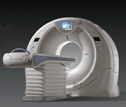
Image: The Aquilion One 320-slice computed tomography (CT) scanner (Photo courtesy of Toshiba Medical Systems).
The world's first dynamic volume computed tomography (CT) scanner has been developed, which can scan a heart in a single heartbeat while administering just one-fifth of the radiation dose of conventional scanners.
Called the Aquilion One, developed by Toshiba Medical Systems (Tokyo, Japan), the new 320-slice CT machine is the first to allow radiologists to view continuous four-dimensional (4D; similar to video) real-time images of the heart and brain without the patient having to move up and down through the scanner.
With its 16-cm detector--five times the size of conventional 64-slice CT scanners--and its dynamic volume CT imaging, clinicians will now be able to observe blood flow (perfusion), movement, and other functions of entire organs, and in precise detail. This coverage of the body will eliminate the need to combine separate scans of organs that fit within the detector area.
Using the Aquilion One, cardiac scanning administers approximately 20% of the radiation dose of a 64-slice conventional CT scanner, and reduces radiation doses by 50% in scans for acute stroke. "Until now, concerns regarding radiation have prevented doctors using more CT to assess the heart and brain,” said Dr. Russell Bull, consultant radiologist at the Royal Bournemouth Hospital (UK). "However, this technology allows very fast and accurate scanning of these organs using much lower radiation doses.”
The new technology will also provide additional benefits for doctors, patients, and hospital managers. The CT system's high-resolution, dynamic volume imaging may reduce the need for multiple scans and invasive procedures, which are frequently more expensive, time-consuming, and less comfortable for the patient.
The system scans extremely quickly, taking just one revolution (0.35 seconds) to scan an entire organ, unlike conventional systems, which revolve many times. For patients presenting with chest pain, the entire heart can be scanned in a single heartbeat. It can also scan one area continuously, providing extremely quick and more precise information on the functionality of an organ. For patients presenting with acute stroke symptoms, just one examination taking no more than 60 seconds can provide critical data on blood flow through the brain including vascular analysis measures. This information could to be key to improving the rapid assessment and treatment of acute stroke.
Other organs can also be seen functioning, such as the lungs as the patient breathes in and out, and joints as the patient flexes them. "With the volume scanning on the Aquilion One, I can see things that I never saw on any 64-slice scanner. This gives me unique diagnostic information,” stated Dr. Patrik Rogalla, chief radiologist and director of the CT division the Charité University Hospital, Berlin (Germany), the first center to install the new scanner in Europe.
The Aquilion One took 10 years to develop and at a cost of US$500 million. The new system has recently been installed in an additional two European university hospital centers. Announcements of UK installations are expected to follow.
Commenting on the European launch of the new scanner, Mark Hitchman, UK general manager of CT Systems at Toshiba Medical Systems, said, "The development of the Aquilion One is a major step-change in the field of CT scanning technology. Its speed of acquisition, coupled with its reduced radiation doses will have a significant impact on the way we use computerized tomography and how we diagnose serious health conditions. It may also open doors to new clinical applications which we never deemed possible.”
Toshiba Medical Systems Corp., an independent group company of Toshiba Corp., is a global leading manufacturer of diagnostic medical imaging systems and comprehensive medical solutions such as CT, cath and electrophysiology (EP) labs, X-ray, ultrasound, nuclear medicine, magnetic resonance imaging (MRI), and information systems.
Related Links:
Toshiba Medical Systems
Latest Radiography News
- AI Improves Early Detection of Interval Breast Cancers
- World's Largest Class Single Crystal Diamond Radiation Detector Opens New Possibilities for Diagnostic Imaging
- AI-Powered Imaging Technique Shows Promise in Evaluating Patients for PCI
- Higher Chest X-Ray Usage Catches Lung Cancer Earlier and Improves Survival
- AI-Powered Mammograms Predict Cardiovascular Risk
- Generative AI Model Significantly Reduces Chest X-Ray Reading Time
- AI-Powered Mammography Screening Boosts Cancer Detection in Single-Reader Settings
- Photon Counting Detectors Promise Fast Color X-Ray Images
- AI Can Flag Mammograms for Supplemental MRI
- 3D CT Imaging from Single X-Ray Projection Reduces Radiation Exposure
- AI Method Accurately Predicts Breast Cancer Risk by Analyzing Multiple Mammograms
- Printable Organic X-Ray Sensors Could Transform Treatment for Cancer Patients
- Highly Sensitive, Foldable Detector to Make X-Rays Safer
- Novel Breast Cancer Screening Technology Could Offer Superior Alternative to Mammogram
- Artificial Intelligence Accurately Predicts Breast Cancer Years Before Diagnosis
- AI-Powered Chest X-Ray Detects Pulmonary Nodules Three Years Before Lung Cancer Symptoms
Channels
MRI
view channel
Cutting-Edge MRI Technology to Revolutionize Diagnosis of Common Heart Problem
Aortic stenosis is a common and potentially life-threatening heart condition. It occurs when the aortic valve, which regulates blood flow from the heart to the rest of the body, becomes stiff and narrow.... Read more
New MRI Technique Reveals True Heart Age to Prevent Attacks and Strokes
Heart disease remains one of the leading causes of death worldwide. Individuals with conditions such as diabetes or obesity often experience accelerated aging of their hearts, sometimes by decades.... Read more
AI Tool Predicts Relapse of Pediatric Brain Cancer from Brain MRI Scans
Many pediatric gliomas are treatable with surgery alone, but relapses can be catastrophic. Predicting which patients are at risk for recurrence remains challenging, leading to frequent follow-ups with... Read more
AI Tool Tracks Effectiveness of Multiple Sclerosis Treatments Using Brain MRI Scans
Multiple sclerosis (MS) is a condition in which the immune system attacks the brain and spinal cord, leading to impairments in movement, sensation, and cognition. Magnetic Resonance Imaging (MRI) markers... Read moreUltrasound
view channel.jpeg)
AI-Powered Lung Ultrasound Outperforms Human Experts in Tuberculosis Diagnosis
Despite global declines in tuberculosis (TB) rates in previous years, the incidence of TB rose by 4.6% from 2020 to 2023. Early screening and rapid diagnosis are essential elements of the World Health... Read more
AI Identifies Heart Valve Disease from Common Imaging Test
Tricuspid regurgitation is a condition where the heart's tricuspid valve does not close completely during contraction, leading to backward blood flow, which can result in heart failure. A new artificial... Read moreNuclear Medicine
view channel
Novel Radiolabeled Antibody Improves Diagnosis and Treatment of Solid Tumors
Interleukin-13 receptor α-2 (IL13Rα2) is a cell surface receptor commonly found in solid tumors such as glioblastoma, melanoma, and breast cancer. It is minimally expressed in normal tissues, making it... Read more
Novel PET Imaging Approach Offers Never-Before-Seen View of Neuroinflammation
COX-2, an enzyme that plays a key role in brain inflammation, can be significantly upregulated by inflammatory stimuli and neuroexcitation. Researchers suggest that COX-2 density in the brain could serve... Read moreGeneral/Advanced Imaging
view channel
AI-Based CT Scan Analysis Predicts Early-Stage Kidney Damage Due to Cancer Treatments
Radioligand therapy, a form of targeted nuclear medicine, has recently gained attention for its potential in treating specific types of tumors. However, one of the potential side effects of this therapy... Read more
CT-Based Deep Learning-Driven Tool to Enhance Liver Cancer Diagnosis
Medical imaging, such as computed tomography (CT) scans, plays a crucial role in oncology, offering essential data for cancer detection, treatment planning, and monitoring of response to therapies.... Read moreImaging IT
view channel
New Google Cloud Medical Imaging Suite Makes Imaging Healthcare Data More Accessible
Medical imaging is a critical tool used to diagnose patients, and there are billions of medical images scanned globally each year. Imaging data accounts for about 90% of all healthcare data1 and, until... Read more
Global AI in Medical Diagnostics Market to Be Driven by Demand for Image Recognition in Radiology
The global artificial intelligence (AI) in medical diagnostics market is expanding with early disease detection being one of its key applications and image recognition becoming a compelling consumer proposition... Read moreIndustry News
view channel
GE HealthCare and NVIDIA Collaboration to Reimagine Diagnostic Imaging
GE HealthCare (Chicago, IL, USA) has entered into a collaboration with NVIDIA (Santa Clara, CA, USA), expanding the existing relationship between the two companies to focus on pioneering innovation in... Read more
Patient-Specific 3D-Printed Phantoms Transform CT Imaging
New research has highlighted how anatomically precise, patient-specific 3D-printed phantoms are proving to be scalable, cost-effective, and efficient tools in the development of new CT scan algorithms... Read more
Siemens and Sectra Collaborate on Enhancing Radiology Workflows
Siemens Healthineers (Forchheim, Germany) and Sectra (Linköping, Sweden) have entered into a collaboration aimed at enhancing radiologists' diagnostic capabilities and, in turn, improving patient care... Read more


