AI Tool Identifies Cancer Type and Changes in Lung Tumor
|
By MedImaging International staff writers Posted on 01 Oct 2018 |

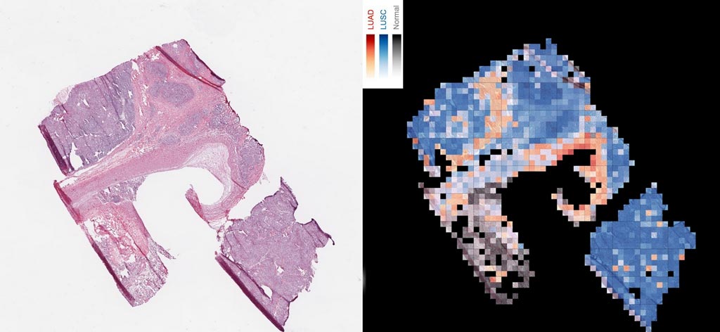
Image: An AI tool analyzes a slice of cancerous tissue to create a map that tells apart two lung cancer types, with squamous cell carcinoma in red, lung squamous cell carcinoma in blue, and normal lung tissue in gray (Photo courtesy of Cision).
Researchers from the NYU School of Medicine (New York City, NY, USA) have developed a new computer program that can analyze the images of patients' lung tumors, specify cancer types, and even identify altered genes driving abnormal cell growth. In their study, the researchers found that the artificial intelligence (AI), or "machine learning," program could distinguish -- with 97% accuracy -- between adenocarcinoma and squamous cell carcinoma, two lung cancer types that experienced pathologists at times struggle to parse without confirmatory tests. Additionally, the study found that the AI was also able to determine from analyzing the images whether the abnormal versions of six genes linked to lung cancer – including EGFR, KRAS and TP53 – were present in cells, with an accuracy ranging from 73% to 86%, depending upon the gene.
For their study, the researchers designed statistical techniques that gave their program the ability to "learn" how to get better at a task, but without being told exactly how. Such programs build rules and mathematical models that enable decision-making based on data examples fed into them, with the program becoming "smarter" as the amount of training data grows.
Newer AI approaches, inspired by nerve cell networks in the brain, use increasingly complex circuits to process information in layers, with each step feeding information into the next, and assigning more or less importance to each piece of information along the way. The researchers trained a deep convolutional neural network, Google's Inception v3, to analyze slide images obtained from The Cancer Genome Atlas, a database of images where cancer diagnoses had already been determined. This allowed the researchers to measure how well their program could be trained to accurately and automatically classify normal versus diseased tissue.
The study found that about half of the small percentage of tumor images misclassified by the study AI program was also misclassified by the pathologists, highlighting the difficulty in distinguishing between the two lung cancer types. On the other hand, 45 out of 54 of the images misclassified by at least one of the pathologists in the study were assigned to the correct cancer type by the machine-learning program, suggesting that AI could offer a useful second opinion.
The researchers now plan to continue training their AI program with data until it can determine which genes are mutated in a given cancer with more than 90% accuracy, after which they will begin seeking government approval to use the technology clinically, and in the diagnosis of several cancer types.
"Delaying the start of cancer treatment is never good," said senior study author Aristotelis Tsirigos, PhD, associate professor in the Department of Pathology at NYU School of Medicine and NYU Langone Health's Perlmutter Cancer Center. "Our study provides strong evidence that an AI approach will be able to instantly determine cancer subtype and mutational profile to get patients started on targeted therapies sooner."
"In our study, we were excited to improve on pathologist-level accuracies, and to show that AI can discover previously unknown patterns in the visible features of cancer cells and the tissues around them," said the study’s co-corresponding author Narges Razavian, PhD, assistant professor in the departments of Radiology and Population Health. "The synergy between data and computational power is creating unprecedented opportunities to improve both the practice and the science of medicine."
Related Links:
NYU School of Medicine
Latest Industry News News
- Hologic Acquires UK-Based Breast Surgical Guidance Company Endomagnetics Ltd.
- Bayer and Google Partner on New AI Product for Radiologists
- Samsung and Bracco Enter Into New Diagnostic Ultrasound Technology Agreement
- IBA Acquires Radcal to Expand Medical Imaging Quality Assurance Offering
- International Societies Suggest Key Considerations for AI Radiology Tools
- Samsung's X-Ray Devices to Be Powered by Lunit AI Solutions for Advanced Chest Screening
- Canon Medical and Olympus Collaborate on Endoscopic Ultrasound Systems
- GE HealthCare Acquires AI Imaging Analysis Company MIM Software
- First Ever International Criteria Lays Foundation for Improved Diagnostic Imaging of Brain Tumors
- RSNA Unveils 10 Most Cited Radiology Studies of 2023
- RSNA 2023 Technical Exhibits to Offer Innovations in AI, 3D Printing and More
- AI Medical Imaging Products to Increase Five-Fold by 2035, Finds Study
- RSNA 2023 Technical Exhibits to Highlight Latest Medical Imaging Innovations
- AI-Powered Technologies to Aid Interpretation of X-Ray and MRI Images for Improved Disease Diagnosis
- Hologic and Bayer Partner to Improve Mammography Imaging
- Global Fixed and Mobile C-Arms Market Driven by Increasing Surgical Procedures
Channels
Radiography
view channel
Novel Breast Imaging System Proves As Effective As Mammography
Breast cancer remains the most frequently diagnosed cancer among women. It is projected that one in eight women will be diagnosed with breast cancer during her lifetime, and one in 42 women who turn 50... Read more
AI Assistance Improves Breast-Cancer Screening by Reducing False Positives
Radiologists typically detect one case of cancer for every 200 mammograms reviewed. However, these evaluations often result in false positives, leading to unnecessary patient recalls for additional testing,... Read moreMRI
view channel
Low-Cost Whole-Body MRI Device Combined with AI Generates High-Quality Results
Magnetic Resonance Imaging (MRI) has significantly transformed healthcare, providing a noninvasive, radiation-free method for detailed imaging. It is especially promising for the future of medical diagnosis... Read more
World's First Whole-Body Ultra-High Field MRI Officially Comes To Market
The world's first whole-body ultra-high field (UHF) MRI has officially come to market, marking a remarkable advancement in diagnostic radiology. United Imaging (Shanghai, China) has secured clearance from the U.... Read moreUltrasound
view channel.jpg)
Diagnostic System Automatically Analyzes TTE Images to Identify Congenital Heart Disease
Congenital heart disease (CHD) is one of the most prevalent congenital anomalies worldwide, presenting substantial health and financial challenges for affected patients. Early detection and treatment of... Read more
Super-Resolution Imaging Technique Could Improve Evaluation of Cardiac Conditions
The heart depends on efficient blood circulation to pump blood throughout the body, delivering oxygen to tissues and removing carbon dioxide and waste. Yet, when heart vessels are damaged, it can disrupt... Read more
First AI-Powered POC Ultrasound Diagnostic Solution Helps Prioritize Cases Based On Severity
Ultrasound scans are essential for identifying and diagnosing various medical conditions, but often, patients must wait weeks or months for results due to a shortage of qualified medical professionals... Read moreNuclear Medicine
view channelNew PET Agent Rapidly and Accurately Visualizes Lesions in Clear Cell Renal Cell Carcinoma Patients
Clear cell renal cell cancer (ccRCC) represents 70-80% of renal cell carcinoma cases. While localized disease can be effectively treated with surgery and ablative therapies, one-third of patients either... Read more
New Imaging Technique Monitors Inflammation Disorders without Radiation Exposure
Imaging inflammation using traditional radiological techniques presents significant challenges, including radiation exposure, poor image quality, high costs, and invasive procedures. Now, new contrast... Read more
New SPECT/CT Technique Could Change Imaging Practices and Increase Patient Access
The development of lead-212 (212Pb)-PSMA–based targeted alpha therapy (TAT) is garnering significant interest in treating patients with metastatic castration-resistant prostate cancer. The imaging of 212Pb,... Read moreGeneral/Advanced Imaging
view channel
Radiation Therapy Computed Tomography Solution Boosts Imaging Accuracy
One of the most significant challenges in oncology care is disease complexity in terms of the variety of cancer types and the individualized presentation of each patient. This complexity necessitates a... Read more
PET Scans Reveal Hidden Inflammation in Multiple Sclerosis Patients
A key challenge for clinicians treating patients with multiple sclerosis (MS) is that after a certain amount of time, they continue to worsen even though their MRIs show no change. A new study has now... Read moreImaging IT
view channel
New Google Cloud Medical Imaging Suite Makes Imaging Healthcare Data More Accessible
Medical imaging is a critical tool used to diagnose patients, and there are billions of medical images scanned globally each year. Imaging data accounts for about 90% of all healthcare data1 and, until... Read more


