Hyperscan Proton Therapy System Offers Pinpoint Accuracy
|
By MedImaging International staff writers Posted on 14 May 2018 |

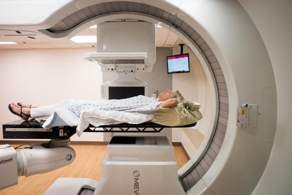
Image: The S250i proton therapy system with Hyperscan PBS (Photo courtesy of Mevion Medial Systems/MedStar Georgetown).
An innovative Proton Therapy System improves on existing scanning capabilities and enables clinicians to deliver conformal fields faster and with more precision than in the past.
The Mevion Medical Systems (Mevion; Littleton, MA, USA) S250i Proton Therapy System with Hyperscan pencil beam scanning (PBS) shapes the delivered radiation dose by precisely targeting tumors with sub-atomic particles. To improve accuracy and speed, a robotically controlled adaptive aperture proton multi-leaf collimator (pMLC) is used, which trims the edges of the beam to achieve sharp lateral dose gradients. This capability can deliver up to a three times sharper drop off in radiation at the delivery field edge, sparing healthy tissue and limiting unnecessary radiation to sensitive locations.
By reducing delivery times to less than five seconds, errors resulting from target tumor shifting under normal organ motion, such as breathing, can be reduced. Other features of the S250i include a gantry mounted superconducting synchrocyclotron, a six degree-of-freedom treatment couch, and advanced in-room image guidance that creates a three-dimensional (3D) representation of the tumor and relays the information to the proton therapy device. The computerized tomography (CT) based imager can also be used to modify treatment in response to changes in the patient’s anatomy, such as tumor shrinkage.
“Proton therapy is an advanced form of radiation that can destroy cancer cells. A machine called a cyclotron speeds up protons to two thirds the speed of light and they become highly charged,” said radiation oncologist Peter Ahn, MD, of MedStar Georgetown (Washington DC, USA), which inaugurated the first system worldwide in April 2018. “Because we can more tightly control the protons than we are able with traditional radiation, proton therapy can be given without damaging critical tissues and structures near the tumor because the beam conforms precisely to the tumor’s size and shape.”
“Delivering sharp field edges has been a real challenge for PBS, especially in shallow fields. In intracranial procedures, where critical structures are in close proximity to tumors at shallow depths, having the sharpest lateral penumbra is essential,” said Skip Rosenthal, VP of clinical education at Mevion. “The sharp penumbras of the adaptive aperture system have substantial benefits for these patients. In addition, the enhanced speed of Hyperscan PBS could enable greater confidence in treating thoracic tumors.”
Proton therapy is a precise form of radiotherapy (RT) that uses charged particles instead of x-rays. It can be a more effective form of treatment than conventional RT as it directs the beam more precisely, with minimal damage to surrounding tissue. Evidence is growing that protons can be effective in treating a number of cancers, in particular children and young people with brain tumors, for whom it appears to produce fewer side effects such as secondary cancers, growth deformity, hearing loss, and learning difficulties.
Related Links:
Mevion Medical Systems
Latest General/Advanced Imaging News
- Imaging Software Improves Lung Diagnosis in Patients Allergic To Medical Contrast Dye
- Bone Density Test Uses Existing CT Images to Predict Fractures
- AI Predicts Cardiac Risk and Mortality from Routine Chest CT Scans
- Radiation Therapy Computed Tomography Solution Boosts Imaging Accuracy
- PET Scans Reveal Hidden Inflammation in Multiple Sclerosis Patients
- Artificial Intelligence Evaluates Cardiovascular Risk from CT Scans
- New AI Method Captures Uncertainty in Medical Images
- CT Coronary Angiography Reduces Need for Invasive Tests to Diagnose Coronary Artery Disease
- Novel Blood Test Could Reduce Need for PET Imaging of Patients with Alzheimer’s
- CT-Based Deep Learning Algorithm Accurately Differentiates Benign From Malignant Vertebral Fractures
- Minimally Invasive Procedure Could Help Patients Avoid Thyroid Surgery
- Self-Driving Mobile C-Arm Reduces Imaging Time during Surgery
- AR Application Turns Medical Scans Into Holograms for Assistance in Surgical Planning
- Imaging Technology Provides Ground-Breaking New Approach for Diagnosing and Treating Bowel Cancer
- CT Coronary Calcium Scoring Predicts Heart Attacks and Strokes
- AI Model Detects 90% of Lymphatic Cancer Cases from PET and CT Images
Channels
Radiography
view channel
Novel Breast Imaging System Proves As Effective As Mammography
Breast cancer remains the most frequently diagnosed cancer among women. It is projected that one in eight women will be diagnosed with breast cancer during her lifetime, and one in 42 women who turn 50... Read more
AI Assistance Improves Breast-Cancer Screening by Reducing False Positives
Radiologists typically detect one case of cancer for every 200 mammograms reviewed. However, these evaluations often result in false positives, leading to unnecessary patient recalls for additional testing,... Read moreMRI
view channel
Low-Cost Whole-Body MRI Device Combined with AI Generates High-Quality Results
Magnetic Resonance Imaging (MRI) has significantly transformed healthcare, providing a noninvasive, radiation-free method for detailed imaging. It is especially promising for the future of medical diagnosis... Read more
World's First Whole-Body Ultra-High Field MRI Officially Comes To Market
The world's first whole-body ultra-high field (UHF) MRI has officially come to market, marking a remarkable advancement in diagnostic radiology. United Imaging (Shanghai, China) has secured clearance from the U.... Read moreUltrasound
view channel
Portable Ultrasound Tool Uses AI to Detect Arm Fractures More Quickly
Suspected injuries to the upper limbs are a major reason for visits to hospital emergency departments. Currently, wait times for an X-ray and subsequent doctor consultation can vary widely, typically ranging... Read more.jpg)
Diagnostic System Automatically Analyzes TTE Images to Identify Congenital Heart Disease
Congenital heart disease (CHD) is one of the most prevalent congenital anomalies worldwide, presenting substantial health and financial challenges for affected patients. Early detection and treatment of... Read more
Super-Resolution Imaging Technique Could Improve Evaluation of Cardiac Conditions
The heart depends on efficient blood circulation to pump blood throughout the body, delivering oxygen to tissues and removing carbon dioxide and waste. Yet, when heart vessels are damaged, it can disrupt... Read more
First AI-Powered POC Ultrasound Diagnostic Solution Helps Prioritize Cases Based On Severity
Ultrasound scans are essential for identifying and diagnosing various medical conditions, but often, patients must wait weeks or months for results due to a shortage of qualified medical professionals... Read moreNuclear Medicine
view channel
New PET Biomarker Predicts Success of Immune Checkpoint Blockade Therapy
Immunotherapies, such as immune checkpoint blockade (ICB), have shown promising clinical results in treating melanoma, non-small cell lung cancer, and other tumor types. However, the effectiveness of these... Read moreNew PET Agent Rapidly and Accurately Visualizes Lesions in Clear Cell Renal Cell Carcinoma Patients
Clear cell renal cell cancer (ccRCC) represents 70-80% of renal cell carcinoma cases. While localized disease can be effectively treated with surgery and ablative therapies, one-third of patients either... Read more
New Imaging Technique Monitors Inflammation Disorders without Radiation Exposure
Imaging inflammation using traditional radiological techniques presents significant challenges, including radiation exposure, poor image quality, high costs, and invasive procedures. Now, new contrast... Read more
New SPECT/CT Technique Could Change Imaging Practices and Increase Patient Access
The development of lead-212 (212Pb)-PSMA–based targeted alpha therapy (TAT) is garnering significant interest in treating patients with metastatic castration-resistant prostate cancer. The imaging of 212Pb,... Read moreImaging IT
view channel
New Google Cloud Medical Imaging Suite Makes Imaging Healthcare Data More Accessible
Medical imaging is a critical tool used to diagnose patients, and there are billions of medical images scanned globally each year. Imaging data accounts for about 90% of all healthcare data1 and, until... Read more
Global AI in Medical Diagnostics Market to Be Driven by Demand for Image Recognition in Radiology
The global artificial intelligence (AI) in medical diagnostics market is expanding with early disease detection being one of its key applications and image recognition becoming a compelling consumer proposition... Read moreIndustry News
view channel
Hologic Acquires UK-Based Breast Surgical Guidance Company Endomagnetics Ltd.
Hologic, Inc. (Marlborough, MA, USA) has entered into a definitive agreement to acquire Endomagnetics Ltd. (Cambridge, UK), a privately held developer of breast cancer surgery technologies, for approximately... Read more
Bayer and Google Partner on New AI Product for Radiologists
Medical imaging data comprises around 90% of all healthcare data, and it is a highly complex and rich clinical data modality and serves as a vital tool for diagnosing patients. Each year, billions of medical... Read more


