Pushing the Boundaries with CEUS and US Elastography
|
By MedImaging International staff writers Posted on 14 Feb 2017 |
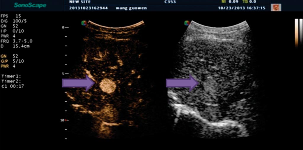
Image: Indeterminate liver lesion in patient with cirrhosis (arrow) demonstrating avid arterial phase hyper-enhancement, suspicious for HCC (Photo courtesy of Andrej Lyshchik, M.D., Ph.D., Thomas Jefferson University Hospital).
Ultrasound elastography and contrast enhanced ultrasound (CEUS) are among the latest advances in ultrasound (US) technology that offer improved spatial and temporal resolution in the detection and characterization of abnormal tissues. SonoScape is leading the field in developing this technology that is improving outcomes for patients.
Leader in the field, Andrej Lyshchik, M.D., Ph.D., Assistant Professor at the Department of Radiology at Thomas Jefferson University Hospital in Philadelphia, US, is enthusiastic for SonoScape’s US elastography. He welcomed the nature of US elastography as a non-invasive technique that allows detection and characterization of tissues with abnormal biomechanical properties.
US Elastography
Currently, there are three main types of US elastography: transient elastography, compression elastography and Acoustic Radiation Force Impulse (ARFI) imaging. Transient elastography uses an external device that generates mechanical displacement of tissues and a built-in US transducer to register shear wave that propagates within the examined tissues. ARFI imaging uses acoustic radiation forces to generate tissue displacement and map its elastic properties based on the speed of sheer wave propagation.
To date, liver, kidney, breast and prostate are among the organs that show most benefit from these technologies. In particular, Dr Lyshchik explained that one of the most commonly used clinical applications of ARFI and transient elastography is for the evaluation of patients with chronic liver disease.
CEUS in the Evaluation Of Tumor Blood Flow
Likewise, CEUS also promises improvements in patient management with its high resolution, real-time visualization of new blood vessels within tumors that increases diagnostic accuracy. CEUS can be used on SonoScape’s scanners.
CEUS uses US contrast agents that are composed of a gas microbubbles, encapsulated by an outer protein or lipid shell. These microbubbles are of a diameter (1-8 µm) that enables passage through the pulmonary capillaries, but restricts the microbubbles to the vasculature, making them excellent intravascular blood pool agents. Unlike contrast agents for MRI and CT, US contrast agents are not nephrotoxic and have no renal contraindications, making them an exceptionally safe to use.
Detecting such changes in tumor vascularity (including blood flow kinetics and microvascular density) is a recognized indicator of treatment response via visualization of the perfusion of US contrast agents. Parameters such as the time required from injection to contrast arrival, rate of US contrast agent inflow, rate of US contrast agent washout, and cumulative US contrast agents signal over time (an indicator of net blood flow) have all been shown to be potentially useful indicators of treatment response. CEUS also provides the opportunity to create 3D parametric maps of tumor perfusion to illustrate the differences in intra-tumoral blood flow kinetics.
Dr Lyshchik explained that within oncology, CEUS can detect changes in tumor vascularity that provide an indicator of treatment response to certain therapies. He remarked that due to the real time nature of ultrasound and the blood pooling properties of US contrast agents, visualization of UCA perfusion provides an indicator of the blood flow kinetics and microvascular density of the tumor.
CEUS of Liver Nodules and Hepatocellular Carcinoma
Of note, CEUS is also a potentially safer, less expensive and more readily available technique for characterizing focal liver nodules in patients at risk for hepatocellular carcinoma, compared to the current clinical standard. This significant cancer, rated as the fifth most common cancer worldwide with an annual incidence of over 550,000, predominantly affects patients with cirrhosis and chronic hepatitis.
But imaging to diagnose hepatocellular carcinoma can be challenging, especially in patients with advanced cirrhosis, in which structural and physiological alterations of the liver can impair detection of the cancer. However, studies of CEUS, which is available on SonoScape’s scanners in this capacity, have demonstrated safety, high specificity and positive predictive value for diagnosis of hepatocellular carcinoma compared to hepatobiliary agent gadoxetate-enhanced MRI.
In an effort to facilitate the clinical use of CEUS, the American College of Radiology recently introduced the CEUS Liver Imaging Reporting and Data System (CEUS LI-RADS), which provides standardisation of CEUS examination and reporting, and allows liver nodule classification based on their likelihood to be hepatocellular carcinoma.
Finally, worth a mention is CEUS-guided biopsy, which is another application of SonoScape’s technology that targets and biopsies lesions, normally invisible or hard to detect, for example, the small nodules of hepatocellular carcinoma on cirrhosis or adenocarcinoma’s areas in the prostate. It can also target viable areas of large, necrotic tumors.
Latest Ultrasound News
- Wireless Chronic Pain Management Device to Reduce Need for Painkillers and Surgery
- New Medical Ultrasound Imaging Technique Enables ICU Bedside Monitoring
- New Incision-Free Technique Halts Growth of Debilitating Brain Lesions
- AI-Powered Lung Ultrasound Outperforms Human Experts in Tuberculosis Diagnosis
- AI Identifies Heart Valve Disease from Common Imaging Test
- Novel Imaging Method Enables Early Diagnosis and Treatment Monitoring of Type 2 Diabetes
- Ultrasound-Based Microscopy Technique to Help Diagnose Small Vessel Diseases
- Smart Ultrasound-Activated Immune Cells Destroy Cancer Cells for Extended Periods
- Tiny Magnetic Robot Takes 3D Scans from Deep Within Body
- High Resolution Ultrasound Speeds Up Prostate Cancer Diagnosis
- World's First Wireless, Handheld, Whole-Body Ultrasound with Single PZT Transducer Makes Imaging More Accessible
- Artificial Intelligence Detects Undiagnosed Liver Disease from Echocardiograms
- Ultrasound Imaging Non-Invasively Tracks Tumor Response to Radiation and Immunotherapy
- AI Improves Detection of Congenital Heart Defects on Routine Prenatal Ultrasounds
- AI Diagnoses Lung Diseases from Ultrasound Videos with 96.57% Accuracy
- New Contrast Agent for Ultrasound Imaging Ensures Affordable and Safer Medical Diagnostics
Channels
Radiography
view channel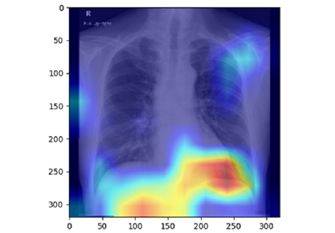
AI Detects Fatty Liver Disease from Chest X-Rays
Fatty liver disease, which results from excess fat accumulation in the liver, is believed to impact approximately one in four individuals globally. If not addressed in time, it can progress to severe conditions... Read more
AI Detects Hidden Heart Disease in Existing CT Chest Scans
Coronary artery calcium (CAC) is a major indicator of cardiovascular risk, but its assessment typically requires a specialized “gated” CT scan that synchronizes with the heartbeat. In contrast, most chest... Read moreMRI
view channel
AI Model Outperforms Doctors at Identifying Patients Most At-Risk of Cardiac Arrest
Hypertrophic cardiomyopathy is one of the most common inherited heart conditions and a leading cause of sudden cardiac death in young individuals and athletes. While many patients live normal lives, some... Read more
New MRI Technique Reveals Hidden Heart Issues
Traditional exercise stress tests conducted within an MRI machine require patients to lie flat, a position that artificially improves heart function by increasing stroke volume due to gravity-driven blood... Read moreNuclear Medicine
view channel
Novel Bacteria-Specific PET Imaging Approach Detects Hard-To-Diagnose Lung Infections
Mycobacteroides abscessus is a rapidly growing mycobacteria that primarily affects immunocompromised patients and those with underlying lung diseases, such as cystic fibrosis or chronic obstructive pulmonary... Read more
New Imaging Approach Could Reduce Need for Biopsies to Monitor Prostate Cancer
Prostate cancer is the second leading cause of cancer-related death among men in the United States. However, the majority of older men diagnosed with prostate cancer have slow-growing, low-risk forms of... Read moreGeneral/Advanced Imaging
view channel
CT Colonography Beats Stool DNA Testing for Colon Cancer Screening
As colorectal cancer remains the second leading cause of cancer-related deaths worldwide, early detection through screening is vital to reduce advanced-stage treatments and associated costs.... Read more
First-Of-Its-Kind Wearable Device Offers Revolutionary Alternative to CT Scans
Currently, patients with conditions such as heart failure, pneumonia, or respiratory distress often require multiple imaging procedures that are intermittent, disruptive, and involve high levels of radiation.... Read more
AI-Based CT Scan Analysis Predicts Early-Stage Kidney Damage Due to Cancer Treatments
Radioligand therapy, a form of targeted nuclear medicine, has recently gained attention for its potential in treating specific types of tumors. However, one of the potential side effects of this therapy... Read moreImaging IT
view channel
New Google Cloud Medical Imaging Suite Makes Imaging Healthcare Data More Accessible
Medical imaging is a critical tool used to diagnose patients, and there are billions of medical images scanned globally each year. Imaging data accounts for about 90% of all healthcare data1 and, until... Read more
Global AI in Medical Diagnostics Market to Be Driven by Demand for Image Recognition in Radiology
The global artificial intelligence (AI) in medical diagnostics market is expanding with early disease detection being one of its key applications and image recognition becoming a compelling consumer proposition... Read moreIndustry News
view channel
GE HealthCare and NVIDIA Collaboration to Reimagine Diagnostic Imaging
GE HealthCare (Chicago, IL, USA) has entered into a collaboration with NVIDIA (Santa Clara, CA, USA), expanding the existing relationship between the two companies to focus on pioneering innovation in... Read more
Patient-Specific 3D-Printed Phantoms Transform CT Imaging
New research has highlighted how anatomically precise, patient-specific 3D-printed phantoms are proving to be scalable, cost-effective, and efficient tools in the development of new CT scan algorithms... Read more
Siemens and Sectra Collaborate on Enhancing Radiology Workflows
Siemens Healthineers (Forchheim, Germany) and Sectra (Linköping, Sweden) have entered into a collaboration aimed at enhancing radiologists' diagnostic capabilities and, in turn, improving patient care... Read more


