PET Scans Help Identify Effective Tuberculosis Drugs
|
By MedImaging International staff writers Posted on 21 Dec 2014 |

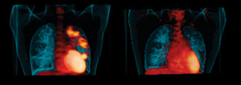
Image: “Hot spots” of infection in a patient’s lungs before treatment (left). Disease improvement after six months of taking the drug linezolid (right) (Photo courtesy of the University of Pittsburgh).
Positron emission tomography (PET) lung imaging can reveal whether or not a treatment drug is able to eradicate tuberculosis (TB) lung infection in human and macaque studies. The new findings indicate that the animal model can effectively predict which experimental drugs have the best likelihood for success in human trials.
In 2012, an estimated 8.6 million people worldwide contracted TB, for which the first-line treatment demands taking four different drugs for six to eight months to get a durable cure, explained senior investigator JoAnne L. Flynn, PhD, professor of microbiology and molecular genetics, University of Pittsburgh (Pitt) School of Medicine (PA, USA). Patients who are not cured of the infection (approximately 500,000 per year) can develop multidrug resistant TB, and have to take as many as six drugs for two years.
The study’s findings were published online December 4, 2014, in the journal Science Translational Medicine. “Some of those people don’t get cured, either, and develop what we call extensively drug-resistant, or XDR, TB, which has a very poor prognosis,” she said. “Our challenge is to find more effective treatments that work in a shorter time period, but the standard preclinical models for testing new drugs have occasionally led to contradictory results when they are evaluated in human trials.”
In an earlier study, Dr. Flynn’s colleagues at the US National Institutes of Health (Bethesda, MD, USA) found that the drug linezolid effectively treated XDR-TB patients who had not improved with conventional treatment, even though mouse studies suggested it would have no impact on the disease. To further examine the effects of linezolid and another drug of the same class, Dr. Flynn and her NIH collaborators, led by Clifton E. Barry III, Ph.D., performed PET/computed tomography (PET/CT) scans in TB-infected humans and macaques, which also get lesions known as granulomas in the lungs. In a PET scan, a tiny amount of a radioactive probe is injected into the blood that gets captured by metabolically active cells, leaving a “hot spot” on the image.
The researchers noted that humans and macaques had very similar disease profiles, and that both groups had hot spots of TB in the lungs that in most cases improved after drug treatment. In addition, CT scans, which show anatomic detail of the lungs, indicated post-treatment improvement. One patient had a hot spot that got worse, and further testing revealed his TB strain was resistant to linezolid.
The findings show that a macaque model and PET scanning can better predict which drugs are likely to be effective in clinical trials, and that could help get new treatments to patients faster, Dr. Flynn said. The scans also could be useful as a way of confirming drug resistance, but aren’t likely to be implemented routinely. “We plan to use this PET scanning strategy to determine why some lesions don’t respond to certain drugs, and to test candidate anti-TB agents,” she said. “This might give us a way of tailoring treatment to individuals.”
Related Links:
University of Pittsburgh School of Medicine
Latest Nuclear Medicine News
- New PET Agent Rapidly and Accurately Visualizes Lesions in Clear Cell Renal Cell Carcinoma Patients
- New Imaging Technique Monitors Inflammation Disorders without Radiation Exposure
- New SPECT/CT Technique Could Change Imaging Practices and Increase Patient Access
- New Radiotheranostic System Detects and Treats Ovarian Cancer Noninvasively
- AI System Automatically and Reliably Detects Cardiac Amyloidosis Using Scintigraphy Imaging
- Early 30-Minute Dynamic FDG-PET Acquisition Could Halve Lung Scan Times
- New Method for Triggering and Imaging Seizures to Help Guide Epilepsy Surgery
- Radioguided Surgery Accurately Detects and Removes Metastatic Lymph Nodes in Prostate Cancer Patients
- New PET Tracer Detects Inflammatory Arthritis Before Symptoms Appear
- Novel PET Tracer Enhances Lesion Detection in Medullary Thyroid Cancer
- Targeted Therapy Delivers Radiation Directly To Cells in Hard-To-Treat Cancers
- New PET Tracer Noninvasively Identifies Cancer Gene Mutation for More Precise Diagnosis
- Algorithm Predicts Prostate Cancer Recurrence in Patients Treated by Radiation Therapy
- Novel PET Imaging Tracer Noninvasively Identifies Cancer Gene Mutation for More Precise Diagnosis
- Ultrafast Laser Technology to Improve Cancer Treatment
- Low-Dose Radiation Therapy Demonstrates Potential for Treatment of Heart Failure
Channels
Radiography
view channel
Novel Breast Imaging System Proves As Effective As Mammography
Breast cancer remains the most frequently diagnosed cancer among women. It is projected that one in eight women will be diagnosed with breast cancer during her lifetime, and one in 42 women who turn 50... Read more
AI Assistance Improves Breast-Cancer Screening by Reducing False Positives
Radiologists typically detect one case of cancer for every 200 mammograms reviewed. However, these evaluations often result in false positives, leading to unnecessary patient recalls for additional testing,... Read moreMRI
view channel
World's First Whole-Body Ultra-High Field MRI Officially Comes To Market
The world's first whole-body ultra-high field (UHF) MRI has officially come to market, marking a remarkable advancement in diagnostic radiology. United Imaging (Shanghai, China) has secured clearance from the U.... Read more
World's First Sensor Detects Errors in MRI Scans Using Laser Light and Gas
MRI scanners are daily tools for doctors and healthcare professionals, providing unparalleled 3D imaging of the brain, vital organs, and soft tissues, far surpassing other imaging technologies in quality.... Read more
Diamond Dust Could Offer New Contrast Agent Option for Future MRI Scans
Gadolinium, a heavy metal used for over three decades as a contrast agent in medical imaging, enhances the clarity of MRI scans by highlighting affected areas. Despite its utility, gadolinium not only... Read more.jpg)
Combining MRI with PSA Testing Improves Clinical Outcomes for Prostate Cancer Patients
Prostate cancer is a leading health concern globally, consistently being one of the most common types of cancer among men and a major cause of cancer-related deaths. In the United States, it is the most... Read moreUltrasound
view channel
First AI-Powered POC Ultrasound Diagnostic Solution Helps Prioritize Cases Based On Severity
Ultrasound scans are essential for identifying and diagnosing various medical conditions, but often, patients must wait weeks or months for results due to a shortage of qualified medical professionals... Read more
Largest Model Trained On Echocardiography Images Assesses Heart Structure and Function
Foundation models represent an exciting frontier in generative artificial intelligence (AI), yet many lack the specialized medical data needed to make them applicable in healthcare settings.... Read more.jpg)
Groundbreaking Technology Enables Precise, Automatic Measurement of Peripheral Blood Vessels
The current standard of care of using angiographic information is often inadequate for accurately assessing vessel size in the estimated 20 million people in the U.S. who suffer from peripheral vascular disease.... Read moreGeneral/Advanced Imaging
view channel
Radiation Therapy Computed Tomography Solution Boosts Imaging Accuracy
One of the most significant challenges in oncology care is disease complexity in terms of the variety of cancer types and the individualized presentation of each patient. This complexity necessitates a... Read more
PET Scans Reveal Hidden Inflammation in Multiple Sclerosis Patients
A key challenge for clinicians treating patients with multiple sclerosis (MS) is that after a certain amount of time, they continue to worsen even though their MRIs show no change. A new study has now... Read moreImaging IT
view channel
New Google Cloud Medical Imaging Suite Makes Imaging Healthcare Data More Accessible
Medical imaging is a critical tool used to diagnose patients, and there are billions of medical images scanned globally each year. Imaging data accounts for about 90% of all healthcare data1 and, until... Read more
Global AI in Medical Diagnostics Market to Be Driven by Demand for Image Recognition in Radiology
The global artificial intelligence (AI) in medical diagnostics market is expanding with early disease detection being one of its key applications and image recognition becoming a compelling consumer proposition... Read moreIndustry News
view channel
Hologic Acquires UK-Based Breast Surgical Guidance Company Endomagnetics Ltd.
Hologic, Inc. (Marlborough, MA, USA) has entered into a definitive agreement to acquire Endomagnetics Ltd. (Cambridge, UK), a privately held developer of breast cancer surgery technologies, for approximately... Read more
Bayer and Google Partner on New AI Product for Radiologists
Medical imaging data comprises around 90% of all healthcare data, and it is a highly complex and rich clinical data modality and serves as a vital tool for diagnosing patients. Each year, billions of medical... Read more


