US Army Hospital Physicians Create Way to Detect Stress Fractures Faster
|
By MedImaging International staff writers Posted on 03 Mar 2013 |

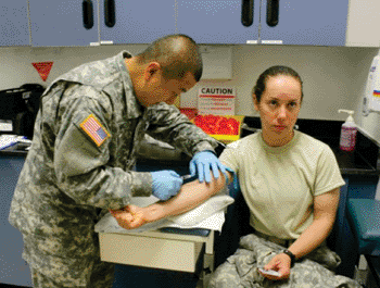
Image: Sgt. Christopher Funke, noncommissioned officer-in-charge of Nuclear Medicine at General Leonard Wood Army Community Hospital, injects Pfc. Sara Reagers, C Company, 84th Chemical Battalion, with a radiopharmaceutical agent (Photo courtesy of John Brooks).

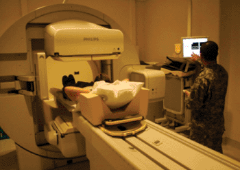
Image: Three to four hours later after the substance has worked its way through the body, the single photon emission computed tomography (SPECT) system can detect the low-level radiation absorbed by stress-injured bone (Photo courtesy of John Brooks).

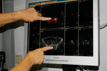
Image: Evaluating the scan (Photo courtesy of John Brooks).
A new imaging protocol developed by the US Army radiologists can now find stress fractures in soldiers much earlier than traditional X-rays.
Single photon emission computed tomography (SPECT) is typically used for nonmusculoskeletal imaging studies, such as cardiac, parathyroid, and adrenal gland imaging. But this ground breaking use of SPECT imaging, in conjunction with a low-dose computed tomography scan, allows General Leonard Wood Army Community Hospital (GLWACH; Lenard Wood, MO, USA) radiologists to optimally rank stress injuries for severity.
With this information, providers return about two thirds more Soldiers back to training after rehabilitation, long before injuries would normally progress to career-ending injuries and incurring large training losses. “SPECT-CT fusion forms a more anatomically-precise image to allow radiologists to pinpoint the exact location of a stress change,” explained Maj. Mustafa Ali-Khan, GLWACH radiology chief. “SPECT-CT is typically not employed in the manner it is here at Fort Leonard Wood. The use of SPECT fusion imaging, although not unique to Army medicine, is unique to its application in its dedicated capacity in musculoskeletal imaging at a Training and Doctrine Command facility, in the manner employed here at Fort Leonard Wood.”
This innovative imaging process was developed in 2010 by the GLWACH radiology department as the result of needs identified through the combined effort of GLWACH staff members in the Musculoskeletal Athletic Team, Orthopedics, Podiatry, and Combined Troop Medical Clinic. “After looking at our training population and our basic training missions, we realized that we could use this technology to our advantage,” Maj. Ali-Khan said.
The technology worked effectively and the numbers are incredible, according to Maj. Ali-Khan. GLWACH’s radiology department used their SPECT-CT fusion imaging technique about 300 times in 2011. “We expect to top 500 studies this year,” Maj. Ali-Khan said.
Typically, injured soldiers in training are given an X-ray and sent back to continue training if an injury is not visible. Missed stress injuries then progress and are discovered at a later date as a more serious insufficiency fracture, or stress fracture. “We have seen a dramatic increase in identification of early stress injuries here at GLWACH, which has led to better outcomes for our Soldier population and less patients who have progressed to more advanced stress injuries and [Medical Evaluation Board] cases,” Maj. Ali-Khan said. “Fewer patients end up with serious stress fractures due to the early intervention. SPECT-CT fusion imaging has worked well in the training environment to mitigate the overall number of bad outcomes with respect to evolving stress injuries. We have every reason to believe that other installations with SPECT equipment, as well as other branches of the military with high training populations subject to bone stress injuries could use our SPECT-CT fusion imaging protocol with a high success rate as well.”
Dr. Ali-Khan reported that the GLWACH medical department activity is fortunate to have this technology, which is typically only available at the Medical Center level at facilities such as Madigan Army Medical Center at Joint Base Lewis-McChord (WA, USA), Walter Reed National Military Medical Center (Bethesda, MD, USA), or Brooke Army Medical Center (San Antonio, TX, USA). “We all feel great to be a part of something so big at such a comparatively small facility,” he said.
It is hard to determine how many training losses will be prevented at GLWACH with SPECT-CT fusion imaging this year, according to Dr. Ali-Khan said. “But we can determine how many stress injuries the technology allows radiologists to catch, and then treat early,” he concluded.
Related Links:
General Leonard Wood Army Community Hospital
Latest MRI News
- World's First Whole-Body Ultra-High Field MRI Officially Comes To Market
- World's First Sensor Detects Errors in MRI Scans Using Laser Light and Gas
- Diamond Dust Could Offer New Contrast Agent Option for Future MRI Scans
- Combining MRI with PSA Testing Improves Clinical Outcomes for Prostate Cancer Patients
- PET/MRI Improves Diagnostic Accuracy for Prostate Cancer Patients
- Next Generation MR-Guided Focused Ultrasound Ushers In Future of Incisionless Neurosurgery
- Two-Part MRI Scan Detects Prostate Cancer More Quickly without Compromising Diagnostic Quality
- World’s Most Powerful MRI Machine Images Living Brain with Unrivaled Clarity
- New Whole-Body Imaging Technology Makes It Possible to View Inflammation on MRI Scan
- Combining Prostate MRI with Blood Test Can Avoid Unnecessary Prostate Biopsies
- New Treatment Combines MRI and Ultrasound to Control Prostate Cancer without Serious Side Effects
- MRI Improves Diagnosis and Treatment of Prostate Cancer
- Combined PET-MRI Scan Improves Treatment for Early Breast Cancer Patients
- 4D MRI Could Improve Clinical Assessment of Heart Blood Flow Abnormalities
- MRI-Guided Focused Ultrasound Therapy Shows Promise in Treating Prostate Cancer
- AI-Based MRI Tool Outperforms Current Brain Tumor Diagnosis Methods
Channels
Radiography
view channel
Novel Breast Imaging System Proves As Effective As Mammography
Breast cancer remains the most frequently diagnosed cancer among women. It is projected that one in eight women will be diagnosed with breast cancer during her lifetime, and one in 42 women who turn 50... Read more
AI Assistance Improves Breast-Cancer Screening by Reducing False Positives
Radiologists typically detect one case of cancer for every 200 mammograms reviewed. However, these evaluations often result in false positives, leading to unnecessary patient recalls for additional testing,... Read moreUltrasound
view channel
First AI-Powered POC Ultrasound Diagnostic Solution Helps Prioritize Cases Based On Severity
Ultrasound scans are essential for identifying and diagnosing various medical conditions, but often, patients must wait weeks or months for results due to a shortage of qualified medical professionals... Read more
Largest Model Trained On Echocardiography Images Assesses Heart Structure and Function
Foundation models represent an exciting frontier in generative artificial intelligence (AI), yet many lack the specialized medical data needed to make them applicable in healthcare settings.... Read more.jpg)
Groundbreaking Technology Enables Precise, Automatic Measurement of Peripheral Blood Vessels
The current standard of care of using angiographic information is often inadequate for accurately assessing vessel size in the estimated 20 million people in the U.S. who suffer from peripheral vascular disease.... Read moreNuclear Medicine
view channelNew PET Agent Rapidly and Accurately Visualizes Lesions in Clear Cell Renal Cell Carcinoma Patients
Clear cell renal cell cancer (ccRCC) represents 70-80% of renal cell carcinoma cases. While localized disease can be effectively treated with surgery and ablative therapies, one-third of patients either... Read more
New Imaging Technique Monitors Inflammation Disorders without Radiation Exposure
Imaging inflammation using traditional radiological techniques presents significant challenges, including radiation exposure, poor image quality, high costs, and invasive procedures. Now, new contrast... Read more
New SPECT/CT Technique Could Change Imaging Practices and Increase Patient Access
The development of lead-212 (212Pb)-PSMA–based targeted alpha therapy (TAT) is garnering significant interest in treating patients with metastatic castration-resistant prostate cancer. The imaging of 212Pb,... Read moreGeneral/Advanced Imaging
view channel
Radiation Therapy Computed Tomography Solution Boosts Imaging Accuracy
One of the most significant challenges in oncology care is disease complexity in terms of the variety of cancer types and the individualized presentation of each patient. This complexity necessitates a... Read more
PET Scans Reveal Hidden Inflammation in Multiple Sclerosis Patients
A key challenge for clinicians treating patients with multiple sclerosis (MS) is that after a certain amount of time, they continue to worsen even though their MRIs show no change. A new study has now... Read moreImaging IT
view channel
New Google Cloud Medical Imaging Suite Makes Imaging Healthcare Data More Accessible
Medical imaging is a critical tool used to diagnose patients, and there are billions of medical images scanned globally each year. Imaging data accounts for about 90% of all healthcare data1 and, until... Read more
Global AI in Medical Diagnostics Market to Be Driven by Demand for Image Recognition in Radiology
The global artificial intelligence (AI) in medical diagnostics market is expanding with early disease detection being one of its key applications and image recognition becoming a compelling consumer proposition... Read moreIndustry News
view channel
Hologic Acquires UK-Based Breast Surgical Guidance Company Endomagnetics Ltd.
Hologic, Inc. (Marlborough, MA, USA) has entered into a definitive agreement to acquire Endomagnetics Ltd. (Cambridge, UK), a privately held developer of breast cancer surgery technologies, for approximately... Read more
Bayer and Google Partner on New AI Product for Radiologists
Medical imaging data comprises around 90% of all healthcare data, and it is a highly complex and rich clinical data modality and serves as a vital tool for diagnosing patients. Each year, billions of medical... Read more


