Smarter CT Scans Could Achieve MRI Diagnostic Capabilities
|
By MedImaging International staff writers Posted on 24 Oct 2023 |

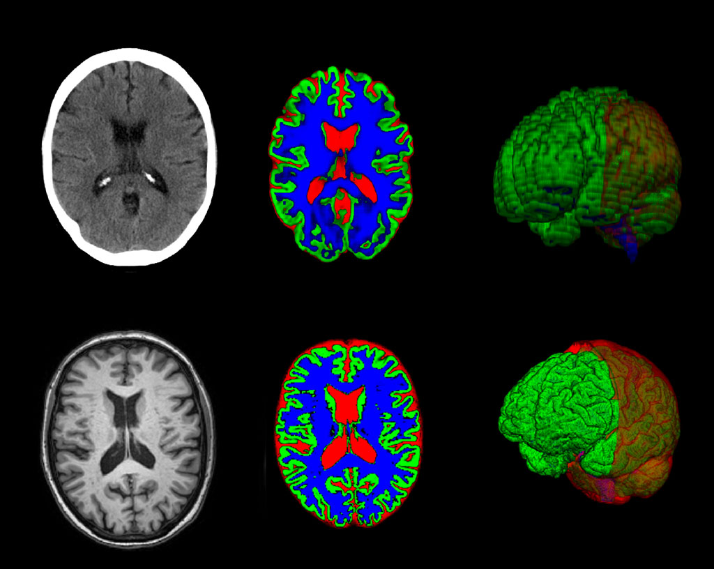
Computed tomography (CT) is a widely used and relatively affordable imaging tool in healthcare, even in areas where other imaging options are scarce. However, when it comes to detailing subtle brain changes or alterations in the ventricular system, CT scans are usually less effective than magnetic resonance imaging (MRI). As a result, specialized imaging often has to be done in bigger hospitals equipped with advanced technology. But now, a novel approach allows CT scans to deliver information on par with MRI in specific situations. This new technique could improve diagnostic capabilities, especially for primary care providers dealing with dementia and other brain disorders.
A new software developed by researchers at the University of Gothenburg (Gothenburg, Sweden) can aid radiologists and other professionals in interpreting CT images. Developed through deep learning, a type of artificial intelligence, the software has been trained on imaging data from 1,117 individuals who had both CT and MRI scans. The software can translate the findings from MRI to CT scans of the same brain, enhancing the clinical application of AI algorithms. The technology aims to serve as a rapid and reliable decision-making tool that minimizes false negatives. It has the potential to refine primary care diagnostics, thereby streamlining the transition of patients to specialized care. In the study, the focus was mainly on healthy older adults and those suffering from different types of dementia.
The team is also exploring the software's application in diagnosing and monitoring normal pressure hydrocephalus (NPH), a condition that affects about 2% of people over the age of 65. NPH, in which fluid builds up in the cerebral ventricular system and results in neurological symptoms, is often challenging to diagnose and can be mistaken for other conditions, which means many cases may go undetected. The software, which has been in development for several years, is undergoing further improvement in collaboration with clinics in Sweden, the UK, and the US, along with a partnering company. This collaboration is essential for the technology to gain approval and integrate into healthcare systems.
“Our method generates diagnostically useful data from routine CT scans that, in some cases, is as good as an MRI scan performed in specialist healthcare,” said Michael Schöll, who led the work involved in the study. “The point is that this simple, quick method can provide much more information from examinations that are already carried out on a routine basis within primary care, but also in certain specialist healthcare investigations. In its initial stage, the method can support dementia diagnosis, however, it is also likely to have other applications within neuroradiology.”
“This is a major step forward for imaging diagnosis,” added Meera Srikrishna, a postdoctor at the University of Gothenburg. “It is now possible to measure the size of different structures or regions of the brain in a similar way to advanced analysis of MRI images. The software makes it possible to segment the brain’s constituent parts in the image and to measure its volume, even though the image quality is not as high with CT.”
Related Links:
University of Gothenburg
Latest General/Advanced Imaging News
- AI-Powered Imaging System Improves Lung Cancer Diagnosis
- AI Model Significantly Enhances Low-Dose CT Capabilities
- Ultra-Low Dose CT Aids Pneumonia Diagnosis in Immunocompromised Patients
- AI Reduces CT Lung Cancer Screening Workload by Almost 80%
- Cutting-Edge Technology Combines Light and Sound for Real-Time Stroke Monitoring
- AI System Detects Subtle Changes in Series of Medical Images Over Time
- New CT Scan Technique to Improve Prognosis and Treatments for Head and Neck Cancers
- World’s First Mobile Whole-Body CT Scanner to Provide Diagnostics at POC
- Comprehensive CT Scans Could Identify Atherosclerosis Among Lung Cancer Patients
- AI Improves Detection of Colorectal Cancer on Routine Abdominopelvic CT Scans
- Super-Resolution Technology Enhances Clinical Bone Imaging to Predict Osteoporotic Fracture Risk
- AI-Powered Abdomen Map Enables Early Cancer Detection
- Deep Learning Model Detects Lung Tumors on CT
- AI Predicts Cardiovascular Risk from CT Scans
- Deep Learning Based Algorithms Improve Tumor Detection in PET/CT Scans
- New Technology Provides Coronary Artery Calcification Scoring on Ungated Chest CT Scans
Channels
Radiography
view channel
World's Largest Class Single Crystal Diamond Radiation Detector Opens New Possibilities for Diagnostic Imaging
Diamonds possess ideal physical properties for radiation detection, such as exceptional thermal and chemical stability along with a quick response time. Made of carbon with an atomic number of six, diamonds... Read more
AI-Powered Imaging Technique Shows Promise in Evaluating Patients for PCI
Percutaneous coronary intervention (PCI), also known as coronary angioplasty, is a minimally invasive procedure where small metal tubes called stents are inserted into partially blocked coronary arteries... Read moreUltrasound
view channel
AI Identifies Heart Valve Disease from Common Imaging Test
Tricuspid regurgitation is a condition where the heart's tricuspid valve does not close completely during contraction, leading to backward blood flow, which can result in heart failure. A new artificial... Read more
Novel Imaging Method Enables Early Diagnosis and Treatment Monitoring of Type 2 Diabetes
Type 2 diabetes is recognized as an autoimmune inflammatory disease, where chronic inflammation leads to alterations in pancreatic islet microvasculature, a key factor in β-cell dysfunction.... Read moreNuclear Medicine
view channel
Novel PET Imaging Approach Offers Never-Before-Seen View of Neuroinflammation
COX-2, an enzyme that plays a key role in brain inflammation, can be significantly upregulated by inflammatory stimuli and neuroexcitation. Researchers suggest that COX-2 density in the brain could serve... Read more
Novel Radiotracer Identifies Biomarker for Triple-Negative Breast Cancer
Triple-negative breast cancer (TNBC), which represents 15-20% of all breast cancer cases, is one of the most aggressive subtypes, with a five-year survival rate of about 40%. Due to its significant heterogeneity... Read moreGeneral/Advanced Imaging
view channel
AI-Powered Imaging System Improves Lung Cancer Diagnosis
Given the need to detect lung cancer at earlier stages, there is an increasing need for a definitive diagnostic pathway for patients with suspicious pulmonary nodules. However, obtaining tissue samples... Read more
AI Model Significantly Enhances Low-Dose CT Capabilities
Lung cancer remains one of the most challenging diseases, making early diagnosis vital for effective treatment. Fortunately, advancements in artificial intelligence (AI) are revolutionizing lung cancer... Read moreImaging IT
view channel
New Google Cloud Medical Imaging Suite Makes Imaging Healthcare Data More Accessible
Medical imaging is a critical tool used to diagnose patients, and there are billions of medical images scanned globally each year. Imaging data accounts for about 90% of all healthcare data1 and, until... Read more
Global AI in Medical Diagnostics Market to Be Driven by Demand for Image Recognition in Radiology
The global artificial intelligence (AI) in medical diagnostics market is expanding with early disease detection being one of its key applications and image recognition becoming a compelling consumer proposition... Read moreIndustry News
view channel
GE HealthCare and NVIDIA Collaboration to Reimagine Diagnostic Imaging
GE HealthCare (Chicago, IL, USA) has entered into a collaboration with NVIDIA (Santa Clara, CA, USA), expanding the existing relationship between the two companies to focus on pioneering innovation in... Read more
Patient-Specific 3D-Printed Phantoms Transform CT Imaging
New research has highlighted how anatomically precise, patient-specific 3D-printed phantoms are proving to be scalable, cost-effective, and efficient tools in the development of new CT scan algorithms... Read more
Siemens and Sectra Collaborate on Enhancing Radiology Workflows
Siemens Healthineers (Forchheim, Germany) and Sectra (Linköping, Sweden) have entered into a collaboration aimed at enhancing radiologists' diagnostic capabilities and, in turn, improving patient care... Read more


