Smaller, Less Expensive Portable MRI Systems to Expand Applications in Various Health Care Settings
|
By MedImaging International staff writers Posted on 19 Jul 2023 |

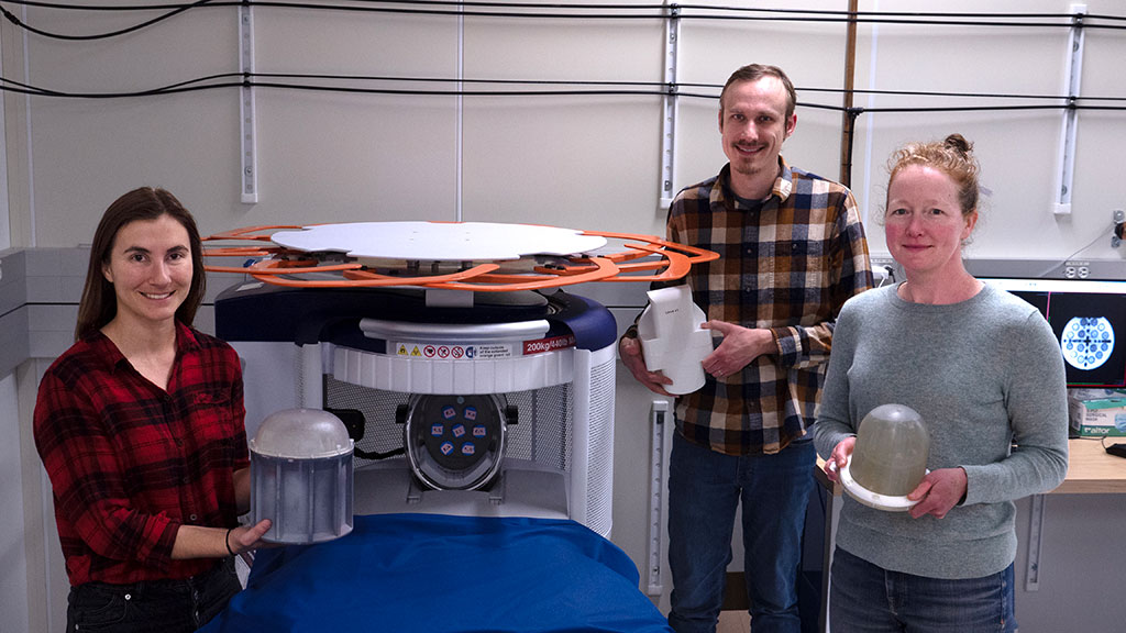
Magnetic Resonance Imaging (MRI) machines offer detailed views of the body's non-bony structures like the brain, muscles, and ligaments, and are instrumental in identifying tumors and diagnosing various ailments. Nonetheless, their high cost, bulkiness, and dependency on powerful magnets limit their availability, primarily to large healthcare facilities. In response to this, companies are designing portable MRI machines that rely on lower-strength magnetic fields. These innovative models hold the potential to extend MRI applications, possibly being incorporated in mobile environments like ambulances. Their reduced cost could also enable greater accessibility, especially in underprivileged communities and developing nations. However, further research is crucial to comprehend the connection between low-field images and the underlying tissue properties they represent.
Researchers at the National Institute of Standards and Technology (NIST, Gaithersburg, MD, USA) have been exploring ways to advance and validate the use of low-field MRI technology for creating images with weaker magnetic fields. In a recent study, the team employed a portable MRI machine available in the market to examine brain tissue characteristics at low magnetic field strength. They used a 64 millitesla magnetic field, significantly lower than traditional MRI machines, to image the brain tissue of ten volunteers. The MRI system was able to produce distinctive images of the entire brain, including its gray matter, white matter, and cerebrospinal fluid. Each of these brain constituents responds uniquely to low magnetic fields, generating distinct signals that offer quantitative information about each component.
Separately, NIST researchers are also investigating materials that could dramatically improve the image quality of low-field MRI scans. MRI contrast agents, which enhance image contrast, making it easier for radiologists to identify anatomical features or evidence of disease, are generally used in MRI at conventional magnetic field strengths but are relatively new in the area of low-field MRI scanners. Researchers have discovered that contrast agents behave differently at lower field strengths, indicating a potential to explore new types of magnetic materials for image enhancement.
The team at NIST tested several magnetic contrast agents' sensitivity at low magnetic fields. The findings revealed that iron oxide nanoparticles were more effective than conventional contrast agents made from gadolinium, a rare-earth metal. At low magnetic field strength, the nanoparticles yielded sufficient contrast utilizing only about one-ninth of the concentration of the gadolinium particles. Moreover, the human body can break down iron oxide nanoparticles, thereby preventing potential accumulation in tissue, unlike gadolinium, which could affect the interpretation of future MRI scans if not accounted for.
Related Links:
NIST
Latest MRI News
- Ultra-Powerful MRI Scans Enable Life-Changing Surgery in Treatment-Resistant Epileptic Patients
- AI-Powered MRI Technology Improves Parkinson’s Diagnoses
- Biparametric MRI Combined with AI Enhances Detection of Clinically Significant Prostate Cancer
- First-Of-Its-Kind AI-Driven Brain Imaging Platform to Better Guide Stroke Treatment Options
- New Model Improves Comparison of MRIs Taken at Different Institutions
- Groundbreaking New Scanner Sees 'Previously Undetectable' Cancer Spread
- First-Of-Its-Kind Tool Analyzes MRI Scans to Measure Brain Aging
- AI-Enhanced MRI Images Make Cancerous Breast Tissue Glow
- AI Model Automatically Segments MRI Images
- New Research Supports Routine Brain MRI Screening in Asymptomatic Late-Stage Breast Cancer Patients
- Revolutionary Portable Device Performs Rapid MRI-Based Stroke Imaging at Patient's Bedside
- AI Predicts After-Effects of Brain Tumor Surgery from MRI Scans
- MRI-First Strategy for Prostate Cancer Detection Proven Safe
- First-Of-Its-Kind 10' x 48' Mobile MRI Scanner Transforms User and Patient Experience
- New Model Makes MRI More Accurate and Reliable
- New Scan Method Shows Effects of Treatment on Lung Function in Real Time
Channels
Radiography
view channel
AI-Powered Imaging Technique Shows Promise in Evaluating Patients for PCI
Percutaneous coronary intervention (PCI), also known as coronary angioplasty, is a minimally invasive procedure where small metal tubes called stents are inserted into partially blocked coronary arteries... Read more
Higher Chest X-Ray Usage Catches Lung Cancer Earlier and Improves Survival
Lung cancer continues to be the leading cause of cancer-related deaths worldwide. While advanced technologies like CT scanners play a crucial role in detecting lung cancer, more accessible and affordable... Read moreUltrasound
view channel
Smart Ultrasound-Activated Immune Cells Destroy Cancer Cells for Extended Periods
Chimeric antigen receptor (CAR) T-cell therapy has emerged as a highly promising cancer treatment, especially for bloodborne cancers like leukemia. This highly personalized therapy involves extracting... Read more
Tiny Magnetic Robot Takes 3D Scans from Deep Within Body
Colorectal cancer ranks as one of the leading causes of cancer-related mortality worldwide. However, when detected early, it is highly treatable. Now, a new minimally invasive technique could significantly... Read more
High Resolution Ultrasound Speeds Up Prostate Cancer Diagnosis
Each year, approximately one million prostate cancer biopsies are conducted across Europe, with similar numbers in the USA and around 100,000 in Canada. Most of these biopsies are performed using MRI images... Read more
World's First Wireless, Handheld, Whole-Body Ultrasound with Single PZT Transducer Makes Imaging More Accessible
Ultrasound devices play a vital role in the medical field, routinely used to examine the body's internal tissues and structures. While advancements have steadily improved ultrasound image quality and processing... Read moreNuclear Medicine
view channel
Novel PET Imaging Approach Offers Never-Before-Seen View of Neuroinflammation
COX-2, an enzyme that plays a key role in brain inflammation, can be significantly upregulated by inflammatory stimuli and neuroexcitation. Researchers suggest that COX-2 density in the brain could serve... Read more
Novel Radiotracer Identifies Biomarker for Triple-Negative Breast Cancer
Triple-negative breast cancer (TNBC), which represents 15-20% of all breast cancer cases, is one of the most aggressive subtypes, with a five-year survival rate of about 40%. Due to its significant heterogeneity... Read moreGeneral/Advanced Imaging
view channel
AI-Powered Imaging System Improves Lung Cancer Diagnosis
Given the need to detect lung cancer at earlier stages, there is an increasing need for a definitive diagnostic pathway for patients with suspicious pulmonary nodules. However, obtaining tissue samples... Read more
AI Model Significantly Enhances Low-Dose CT Capabilities
Lung cancer remains one of the most challenging diseases, making early diagnosis vital for effective treatment. Fortunately, advancements in artificial intelligence (AI) are revolutionizing lung cancer... Read moreImaging IT
view channel
New Google Cloud Medical Imaging Suite Makes Imaging Healthcare Data More Accessible
Medical imaging is a critical tool used to diagnose patients, and there are billions of medical images scanned globally each year. Imaging data accounts for about 90% of all healthcare data1 and, until... Read more
Global AI in Medical Diagnostics Market to Be Driven by Demand for Image Recognition in Radiology
The global artificial intelligence (AI) in medical diagnostics market is expanding with early disease detection being one of its key applications and image recognition becoming a compelling consumer proposition... Read moreIndustry News
view channel
GE HealthCare and NVIDIA Collaboration to Reimagine Diagnostic Imaging
GE HealthCare (Chicago, IL, USA) has entered into a collaboration with NVIDIA (Santa Clara, CA, USA), expanding the existing relationship between the two companies to focus on pioneering innovation in... Read more
Patient-Specific 3D-Printed Phantoms Transform CT Imaging
New research has highlighted how anatomically precise, patient-specific 3D-printed phantoms are proving to be scalable, cost-effective, and efficient tools in the development of new CT scan algorithms... Read more
Siemens and Sectra Collaborate on Enhancing Radiology Workflows
Siemens Healthineers (Forchheim, Germany) and Sectra (Linköping, Sweden) have entered into a collaboration aimed at enhancing radiologists' diagnostic capabilities and, in turn, improving patient care... Read more


