AI Identifies Noncancerous Thyroid Nodules on Ultrasound Images and Reduces Biopsies
|
By MedImaging International staff writers Posted on 13 Jun 2022 |


|
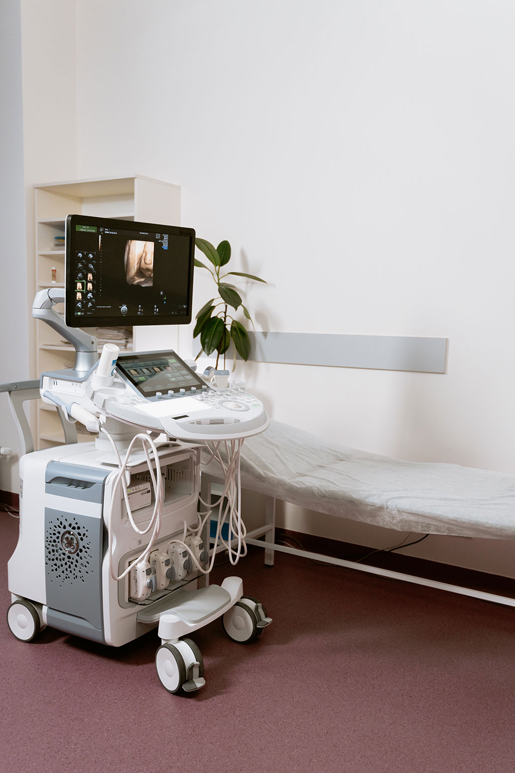
Thyroid nodules are very common. Fine needle aspiration biopsy is used to diagnose thyroid cancer. However most biopsies produce benign (non-cancerous) results and are potentially avoidable. Now, a new study has found that artificial intelligence (AI) can be used to identify thyroid nodules seen on thyroid ultrasound that are very unlikely to be cancerous, reducing a large number of unnecessary biopsies.
In the new study, researchers at the University of Colorado Anschutz Medical Campus (Aurora, CO, USA) used machine learning, a type of AI, to analyze ultrasound images of thyroid nodules. Machine learning is the process of using mathematical models of data to help a computer learn without direct instruction. More than 30,000 images from 621 thyroid nodules were used to train the machine-learning model that classifies thyroid nodules as “cancer” or “no cancer.” The model was tested on a different set of 145 nodules collected at another healthcare system. The AI-based model achieved a sensitivity (ability to not miss cancer) of 97%, and a specificity (ability to correctly identify a cancer) of 61%.
“This study demonstrates that the ultrasound-based AI classifier of thyroid nodules achieves sensitivity comparable to that of thyroid biopsy with fine needle aspiration,” said study lead researcher Nikita Pozdeyev, M.D., Ph.D., of the University of Colorado Anschutz Medical Campus.
“We believe this is a good next step to improving patient care and avoiding unnecessary procedures,” he said. He noted that prospective clinical trials are needed before this tool can be accepted as a standard of care.
“We demonstrated that using AI analysis of ultrasound images to rule out thyroid cancer and avoid biopsy is definitely possible,” he said. “This technology could assist radiologists and endocrinologists in choosing which thyroid nodules should undergo biopsy, especially those in the community who may not review a large number of thyroid ultrasound images.”
Related Links:
University of Colorado Anschutz Medical Campus





Latest Ultrasound News
- Simple 2D Ultrasound Tool Accurately Estimates Placental Volume
- Portable 3D Ultrasound System Enables Reproducible Breast Cancer Monitoring
- AI Ultrasound Platform Receives CE Mark for Prenatal Screening
- Hybrid Imaging Platform Reveals How Sleep Supports Brain Waste Removal
- AI Robotic Ultrasound System Automates Echocardiography and Improves Consistency
- Whole Cross-Section Ultrasound System Enables Operator-Independent Imaging
- New Ultrasound AI Tool Supports Rapid Prenatal Assessment
- New Consensus Standardizes Ultrasound-Based Fatty Liver Assessment
- Groundbreaking Technology to Enhance Precision in Emergency and Critical Care
- Reusable Gel Pad Made from Tamarind Seed Could Transform Ultrasound Examinations
- AI Model Accurately Detects Placenta Accreta in Pregnancy Before Delivery
- Portable Ultrasound Sensor to Enable Earlier Breast Cancer Detection
- Portable Imaging Scanner to Diagnose Lymphatic Disease in Real Time
- Imaging Technique Generates Simultaneous 3D Color Images of Soft-Tissue Structure and Vasculature
- Wearable Ultrasound Imaging System to Enable Real-Time Disease Monitoring
- Ultrasound Technique Visualizes Deep Blood Vessels in 3D Without Contrast Agents
Channels
Radiography
view channel
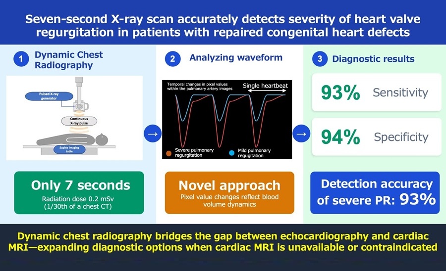
Computational X-Ray Technique Enables High-Resolution, Low-Dose Radiography
Reducing radiation in medical X-ray imaging remains a persistent challenge because dose reductions often degrade spatial resolution and diagnostic confidence. This is especially critical in pediatrics,... Read more
AI Tool Predicts Five-Year Breast Cancer Risk from Mammograms
Breast cancer risk assessment during routine screening is difficult because many women who develop the disease have no known genetic mutations or family history. Static risk tools provide limited discrimination... Read more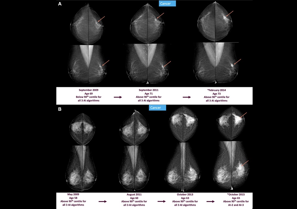
MRI
view channel
Advanced MRI Reveals Structural Brain Changes Linked to Better Cognitive Health
Objective, noninvasive measures that capture subtle, training-related changes in adult brain structure remain limited in routine clinical practice. Quantifying neuroplasticity with precision is especially... Read more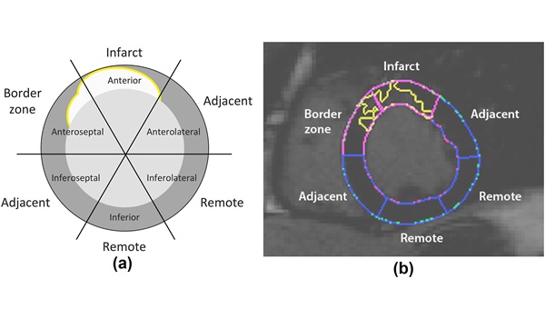
Cardiac MRI Detects Hidden Heart Dysfunction After Heart Attack
Myocardial infarction often leaves patients with persistent ventricular dysfunction that can progress without obvious signs. Detecting early decline remains difficult because standard measures may miss... Read more
Nuclear Medicine
view channel
Radiotherapy Professionals Embrace AI as a Clinical Support Tool
Radiotherapy planning requires precise identification of organs at risk near a tumor, but manual contouring is repetitive and time-consuming, often delaying plan completion and straining specialist capacity.... Read more
Second PSMA PET Identifies Disease in More Than Half of Negative Cases
Biochemical recurrence of prostate cancer after prostatectomy or radiation can be difficult to localize when imaging is negative. Uncertain disease sites may delay salvage therapy and expose patients to... Read more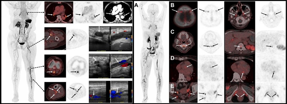
![Image: (A-C) Representative whole-body maximum-intensity projection images and regional fused PET/CT images from three histologically confirmed osteosarcoma patients who underwent paired [68Ga]Ga-B7-H3-BCH PET/CT and 18F-FDGE PET/CT within 7 days. (D) Multimodal imaging evaluation of patient 3, including X-ray, MRI (T2-weighted imaging, T2WI), CT, and B7-H3 PET/CT. (Photo courtesy of SNMMI)](https://globetechcdn.com/mobile_medicalimaging/images/stories/articles/article_images/2026-06-07/294811110_image_1.jpg "Image: (A-C) Representative whole-body maximum-intensity projection images and regional fused PET/CT images from three histologically confirmed osteosarcoma patients who underwent paired [68Ga]Ga-B7-H3-BCH PET/CT and 18F-FDGE PET/CT within 7 days. (D) Multimodal imaging evaluation of patient 3, including X-ray, MRI (T2-weighted imaging, T2WI), CT, and B7-H3 PET/CT. (Photo courtesy of SNMMI)")
General/Advanced Imaging
view channel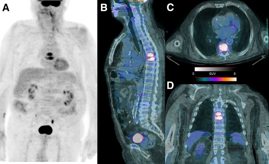
PET/CT Improves Diagnosis and Management of Fever of Unknown Origin
Fever of unknown origin (FUO) is persistent fever without an identifiable cause and remains difficult to diagnose because potential etiologies are broad and no single gold-standard pathway exists.... Read more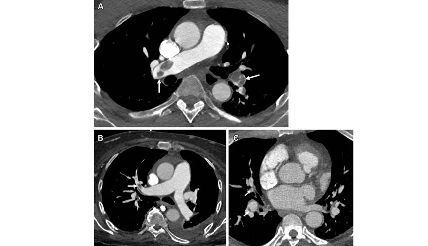
New PE-RADS Framework Standardizes Pulmonary Embolism Imaging Reports
Pulmonary embolism is a sudden blockage of the lung’s blood vessels that reduces pulmonary blood flow, can strain the right hear,t and may be fatal if untreated. Patients often present with shortness of... Read more
Imaging IT
view channel
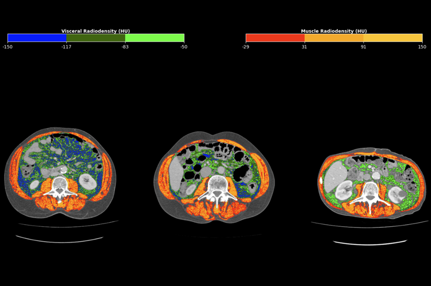
Ambient AI Reporting Platform Streamlines Radiology Reporting
Radiology departments face growing imaging volumes and staffing shortages, creating reporting bottlenecks and pressure to maintain turnaround times. Conventional dictation tools document findings after... Read more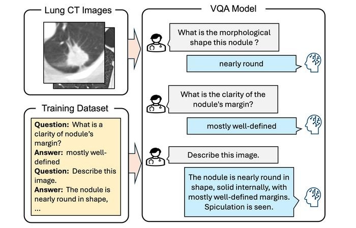
Interactive AI Tool Supports Explainable Lung Nodule Assessment
Lung cancer is a leading cause of cancer mortality, and timely characterization of pulmonary nodules on chest computed tomography (CT) is essential for directing care. Interpreting nodule morphology demands... Read more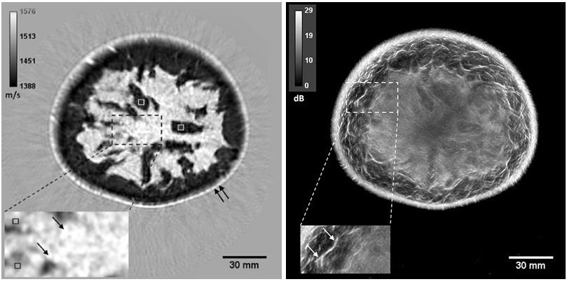
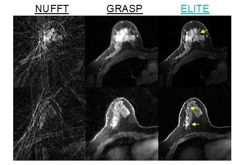
Breast Imaging Software Enhances Visualization and Tissue Characterization in Challenging Cases
Breast imaging can be particularly challenging in cases involving small breasts or implants, where image reconstruction and tissue characterization may be limited. Clinicians also need reproducible analysis... Read more
Industry News
view channel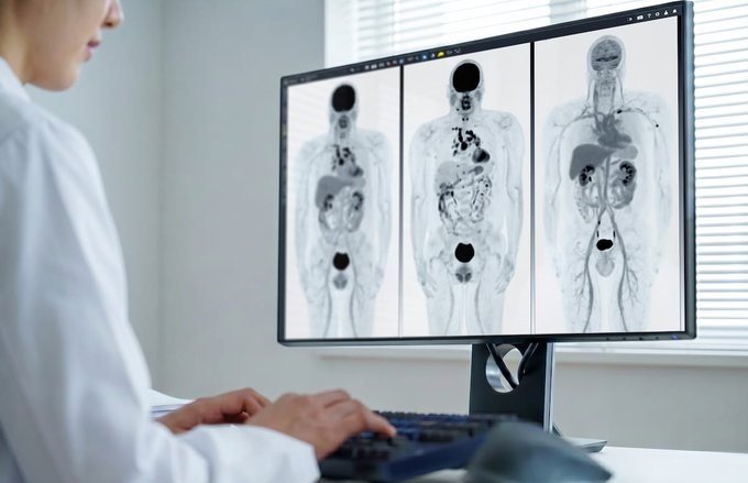
GE HealthCare Showcases AI-Enabled Nuclear Medicine Portfolio at SNMMI 2026
Nuclear medicine is expanding rapidly as health systems adopt theranostics and broaden access to radiopharmaceuticals, increasing demand for scalable operations and consistent diagnostic confidence.... Read more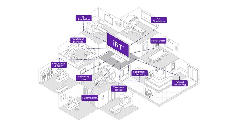
GE HealthCare Highlights AI-Supported Radiation Therapy Tools at ESTRO 2026
At the European Society for Radiotherapy and Oncology (ESTRO) 2026 Congress in Stockholm, GE HealthCare is highlighting Intelligent Radiation Therapy (iRT), MIM Software innovations, and BK Medical surgical... Read more