Cutting-Edge Imaging Pinpoints Where and When Hemorrhagic Stroke Has Occurred
|
By MedImaging International staff writers Posted on 13 Jun 2022 |

Hemorrhagic stroke, where a weakened vessel in the brain ruptures, can lead to permanent disability or death. Across the globe, over 15 million people are coping with its effects. Time is of the essence when it comes to stroke; the sooner doctors can start treatment, the better the odds they can limit damage. Now, a new study has moved us one step closer to identifying when the bleeding associated with a hemorrhagic stroke starts - critical information for improving patient outcomes.
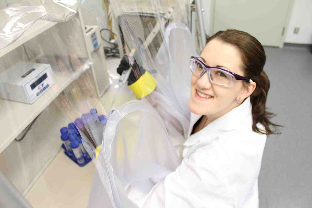
Using the Mid-IR beamline at the Canadian Light Source (CLS) at the University of Saskatchewan (USask, Saskatoon, Canada), the research team examined brain tissue samples with a special technique called Fourier-transform infrared imaging. The novel approach enabled the researchers to identify changes in the brain specific to hemorrhagic stroke. According to the researchers, the combination of the beamline and infrared imaging made it easy to detect markers of brain damage caused by hemorrhagic stroke.
With synchrotron technology, the team could see where a bleed originated and the extent of oxidative damage it caused – something impossible to do with a microscope or traditional approaches to imaging. Armed with this new approach, and a better understanding of what they are looking for, the researchers will now go back through their extensive “library” of stroke tissue samples to gain a clearer picture of the speed at which oxidative damage begins to ramp up. The team’s findings could eventually enable doctors to use clinical imaging – such as MRI or CT scans – to pinpoint where, and how long ago, a hemorrhagic stroke occurred in the brain. Knowing when bleeding has started can provide clinicians with a clearer picture of the time window they have to act.
“In a sense, this is giving us ‘superhuman vision’ to look at these brains and map out what’s happening metabolically,” said Dr. Jake Pushie, a member of the research team at USask’s College of Medicine.
“Being able to understand what is going on biologically, when we see any kinds of changes in the clinical images, could help doctors provide better care when it comes to minimizing the tissue damage associated with stroke,” added Miranda Messmer, another member of the research team.
Related Links:
University of Saskatchewan
Latest Critical Care News
Channels
Radiography
view channel
AI Detects Early Signs of Aging from Chest X-Rays
Chronological age does not always reflect how fast the body is truly aging, and current biological age tests often rely on DNA-based markers that may miss early organ-level decline. Detecting subtle, age-related... Read more
X-Ray Breakthrough Captures Three Image-Contrast Types in Single Shot
Detecting early-stage cancer or subtle changes deep inside tissues has long challenged conventional X-ray systems, which rely only on how structures absorb radiation. This limitation keeps many microstructural... Read moreMRI
view channel
Novel Imaging Approach to Improve Treatment for Spinal Cord Injuries
Vascular dysfunction in the spinal cord contributes to multiple neurological conditions, including traumatic injuries and degenerative cervical myelopathy, where reduced blood flow can lead to progressive... Read more
AI-Assisted Model Enhances MRI Heart Scans
A cardiac MRI can reveal critical information about the heart’s function and any abnormalities, but traditional scans take 30 to 90 minutes and often suffer from poor image quality due to patient movement.... Read more
AI Model Outperforms Doctors at Identifying Patients Most At-Risk of Cardiac Arrest
Hypertrophic cardiomyopathy is one of the most common inherited heart conditions and a leading cause of sudden cardiac death in young individuals and athletes. While many patients live normal lives, some... Read moreUltrasound
view channel
Wearable Ultrasound Imaging System to Enable Real-Time Disease Monitoring
Chronic conditions such as hypertension and heart failure require close monitoring, yet today’s ultrasound imaging is largely confined to hospitals and short, episodic scans. This reactive model limits... Read more
Ultrasound Technique Visualizes Deep Blood Vessels in 3D Without Contrast Agents
Producing clear 3D images of deep blood vessels has long been difficult without relying on contrast agents, CT scans, or MRI. Standard ultrasound typically provides only 2D cross-sections, limiting clinicians’... Read moreNuclear Medicine
view channel
PET Imaging of Inflammation Predicts Recovery and Guides Therapy After Heart Attack
Acute myocardial infarction can trigger lasting heart damage, yet clinicians still lack reliable tools to identify which patients will regain function and which may develop heart failure.... Read more
Radiotheranostic Approach Detects, Kills and Reprograms Aggressive Cancers
Aggressive cancers such as osteosarcoma and glioblastoma often resist standard therapies, thrive in hostile tumor environments, and recur despite surgery, radiation, or chemotherapy. These tumors also... Read more
New Imaging Solution Improves Survival for Patients with Recurring Prostate Cancer
Detecting recurrent prostate cancer remains one of the most difficult challenges in oncology, as standard imaging methods such as bone scans and CT scans often fail to accurately locate small or early-stage tumors.... Read moreGeneral/Advanced Imaging
view channel
New Algorithm Dramatically Speeds Up Stroke Detection Scans
When patients arrive at emergency rooms with stroke symptoms, clinicians must rapidly determine whether the cause is a blood clot or a brain bleed, as treatment decisions depend on this distinction.... Read more
3D Scanning Approach Enables Ultra-Precise Brain Surgery
Precise navigation is critical in neurosurgery, yet even small alignment errors can affect outcomes when operating deep within the brain. A new 3D surface-scanning approach now provides a radiation-free... Read moreImaging IT
view channel
New Google Cloud Medical Imaging Suite Makes Imaging Healthcare Data More Accessible
Medical imaging is a critical tool used to diagnose patients, and there are billions of medical images scanned globally each year. Imaging data accounts for about 90% of all healthcare data1 and, until... Read more
Global AI in Medical Diagnostics Market to Be Driven by Demand for Image Recognition in Radiology
The global artificial intelligence (AI) in medical diagnostics market is expanding with early disease detection being one of its key applications and image recognition becoming a compelling consumer proposition... Read moreIndustry News
view channel
GE HealthCare and NVIDIA Collaboration to Reimagine Diagnostic Imaging
GE HealthCare (Chicago, IL, USA) has entered into a collaboration with NVIDIA (Santa Clara, CA, USA), expanding the existing relationship between the two companies to focus on pioneering innovation in... Read more
Patient-Specific 3D-Printed Phantoms Transform CT Imaging
New research has highlighted how anatomically precise, patient-specific 3D-printed phantoms are proving to be scalable, cost-effective, and efficient tools in the development of new CT scan algorithms... Read more
Siemens and Sectra Collaborate on Enhancing Radiology Workflows
Siemens Healthineers (Forchheim, Germany) and Sectra (Linköping, Sweden) have entered into a collaboration aimed at enhancing radiologists' diagnostic capabilities and, in turn, improving patient care... Read more


 Guided Devices.jpg)


