Fujifilm’s AI Technology Predicts Alzheimer’s Disease Progression Using MRI Images
|
By MedImaging International staff writers Posted on 22 Apr 2022 |

In the development of new drugs for Alzheimer’s disease (AD) in recent years, many clinical trials of patients with mild cognitive impairment (MCI) have been conducted to observe amyloid-β presence, which is the major causal substance of AD and begins accumulating prior to onset of AD. However, most clinical trials have not been successful, and it is difficult to prove statistically significant differences. One of the reasons is that the percentage of patients who progress from MCI to AD within two years is less than 20%, and many MCI patients remain unchanged even if they receive placebo. Now, a new artificial intelligence (AI) technology has been shown to predict the progression from MCI to AD with a high accuracy, even with limited learning data.
FUJIFILM Corporation (Tokyo, Japan) has announced positive study results of its new AI Technology for AD Progression Prediction to predict whether patients with MCI will progress to AD within two years. The AI Technology for AD Progression Prediction with an accuracy rate of 88% was developed by Fujifilm based on its advanced image recognition technologies and machine learning expertise. Numerous research has been reported in recent years indicating that the accuracy of image recognition is enhanced by deep learning technology. In addition, accurate predictions require a large dataset of images, although the open database of NA-ADNI, the world’s largest AD research project, only has images of about 1,000 MCI patients. Generally, establishing deep learning technology requires over 10 million images in the research of object recognition. To overcome this limitation, Fujifilm has built AI Technology for AD Progression Prediction, by targeting specific areas inside the brain that are strongly correlated with the progression of AD.
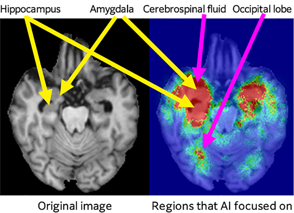
Fujifilm used its advanced image recognition technologies accumulated in the fields of photography and healthcare, and focused on the hippocampus and the anterior temporal lobe, respectively, identified the regions from the three-dimensional MRI brain images that are considered to be strongly correlated with the progression of AD. The company used deep learning to extract detailed atrophy patterns from the described two regions, and calculated them as the image features. AI focused even more on the atrophy patterns in the hippocampus and the amygdala regions which are important regions for AD diagnosis in radiographic image interpretation, and then predicted the progression to AD from those patterns. Fujifilm evaluated the technology’s prediction accuracy by applying the AI Technology for AD Progression Prediction to the databases not only of NA-ADNI but also of J-ADNI that is completely unknown to AI. The findings revealed that the technology’s accuracy in predicting whether patients would progress to AD from MCI was 88% for NA-ADNI and 84% for J-ADNI. AUC, which is an important evaluation index of AI, was 0.95 for NA ADNI and 0.91 for J-ADNI.
The findings verify that the AI Technology for AD Progression Prediction has a high generalizability and can predict which patients would progress from MCI to AD with a high accuracy, even for subjects from different cohorts. Fujifilm will apply the AI technology to the patient stratification, using its prediction results on the clinical trial data, and verify the technology’s usefulness even further. Specifically, it will predict the speed of the patients’ progression to AD, and investigate the possibility of improving the clinical trial’s success rate by excluding the patients who do not progress to AD from the clinical trial, and reducing the gap in the distribution of progression speed between the control group and the treatment group. Moreover, Fujifilm will aim to apply the AI Technology for new clinical trials prospectively. It will also apply the algorithm of the AI Technology to the brain images and clinical data of diverse mental and neural diseases. Fujifilm expects these activities to lead to predicting the prognosis and response to treatment, and can play an important role in promoting personalized medicine.
Related Links:
FUJIFILM Corporation
Latest MRI News
- AI Tool Predicts Relapse of Pediatric Brain Cancer from Brain MRI Scans
- AI Tool Tracks Effectiveness of Multiple Sclerosis Treatments Using Brain MRI Scans
- Ultra-Powerful MRI Scans Enable Life-Changing Surgery in Treatment-Resistant Epileptic Patients
- AI-Powered MRI Technology Improves Parkinson’s Diagnoses
- Biparametric MRI Combined with AI Enhances Detection of Clinically Significant Prostate Cancer
- First-Of-Its-Kind AI-Driven Brain Imaging Platform to Better Guide Stroke Treatment Options
- New Model Improves Comparison of MRIs Taken at Different Institutions
- Groundbreaking New Scanner Sees 'Previously Undetectable' Cancer Spread
- First-Of-Its-Kind Tool Analyzes MRI Scans to Measure Brain Aging
- AI-Enhanced MRI Images Make Cancerous Breast Tissue Glow
- AI Model Automatically Segments MRI Images
- New Research Supports Routine Brain MRI Screening in Asymptomatic Late-Stage Breast Cancer Patients
- Revolutionary Portable Device Performs Rapid MRI-Based Stroke Imaging at Patient's Bedside
- AI Predicts After-Effects of Brain Tumor Surgery from MRI Scans
- MRI-First Strategy for Prostate Cancer Detection Proven Safe
- First-Of-Its-Kind 10' x 48' Mobile MRI Scanner Transforms User and Patient Experience
Channels
Radiography
view channel
World's Largest Class Single Crystal Diamond Radiation Detector Opens New Possibilities for Diagnostic Imaging
Diamonds possess ideal physical properties for radiation detection, such as exceptional thermal and chemical stability along with a quick response time. Made of carbon with an atomic number of six, diamonds... Read more
AI-Powered Imaging Technique Shows Promise in Evaluating Patients for PCI
Percutaneous coronary intervention (PCI), also known as coronary angioplasty, is a minimally invasive procedure where small metal tubes called stents are inserted into partially blocked coronary arteries... Read moreUltrasound
view channel.jpeg)
AI-Powered Lung Ultrasound Outperforms Human Experts in Tuberculosis Diagnosis
Despite global declines in tuberculosis (TB) rates in previous years, the incidence of TB rose by 4.6% from 2020 to 2023. Early screening and rapid diagnosis are essential elements of the World Health... Read more
AI Identifies Heart Valve Disease from Common Imaging Test
Tricuspid regurgitation is a condition where the heart's tricuspid valve does not close completely during contraction, leading to backward blood flow, which can result in heart failure. A new artificial... Read moreNuclear Medicine
view channel
Novel Radiolabeled Antibody Improves Diagnosis and Treatment of Solid Tumors
Interleukin-13 receptor α-2 (IL13Rα2) is a cell surface receptor commonly found in solid tumors such as glioblastoma, melanoma, and breast cancer. It is minimally expressed in normal tissues, making it... Read more
Novel PET Imaging Approach Offers Never-Before-Seen View of Neuroinflammation
COX-2, an enzyme that plays a key role in brain inflammation, can be significantly upregulated by inflammatory stimuli and neuroexcitation. Researchers suggest that COX-2 density in the brain could serve... Read moreGeneral/Advanced Imaging
view channel
AI-Powered Imaging System Improves Lung Cancer Diagnosis
Given the need to detect lung cancer at earlier stages, there is an increasing need for a definitive diagnostic pathway for patients with suspicious pulmonary nodules. However, obtaining tissue samples... Read more
AI Model Significantly Enhances Low-Dose CT Capabilities
Lung cancer remains one of the most challenging diseases, making early diagnosis vital for effective treatment. Fortunately, advancements in artificial intelligence (AI) are revolutionizing lung cancer... Read moreImaging IT
view channel
New Google Cloud Medical Imaging Suite Makes Imaging Healthcare Data More Accessible
Medical imaging is a critical tool used to diagnose patients, and there are billions of medical images scanned globally each year. Imaging data accounts for about 90% of all healthcare data1 and, until... Read more
Global AI in Medical Diagnostics Market to Be Driven by Demand for Image Recognition in Radiology
The global artificial intelligence (AI) in medical diagnostics market is expanding with early disease detection being one of its key applications and image recognition becoming a compelling consumer proposition... Read moreIndustry News
view channel
GE HealthCare and NVIDIA Collaboration to Reimagine Diagnostic Imaging
GE HealthCare (Chicago, IL, USA) has entered into a collaboration with NVIDIA (Santa Clara, CA, USA), expanding the existing relationship between the two companies to focus on pioneering innovation in... Read more
Patient-Specific 3D-Printed Phantoms Transform CT Imaging
New research has highlighted how anatomically precise, patient-specific 3D-printed phantoms are proving to be scalable, cost-effective, and efficient tools in the development of new CT scan algorithms... Read more
Siemens and Sectra Collaborate on Enhancing Radiology Workflows
Siemens Healthineers (Forchheim, Germany) and Sectra (Linköping, Sweden) have entered into a collaboration aimed at enhancing radiologists' diagnostic capabilities and, in turn, improving patient care... Read more


