AI-Enhanced Cardiac MRI Platform Evaluates Congenital Heart Disease in Real Time
|
By MedImaging International staff writers Posted on 08 Apr 2022 |

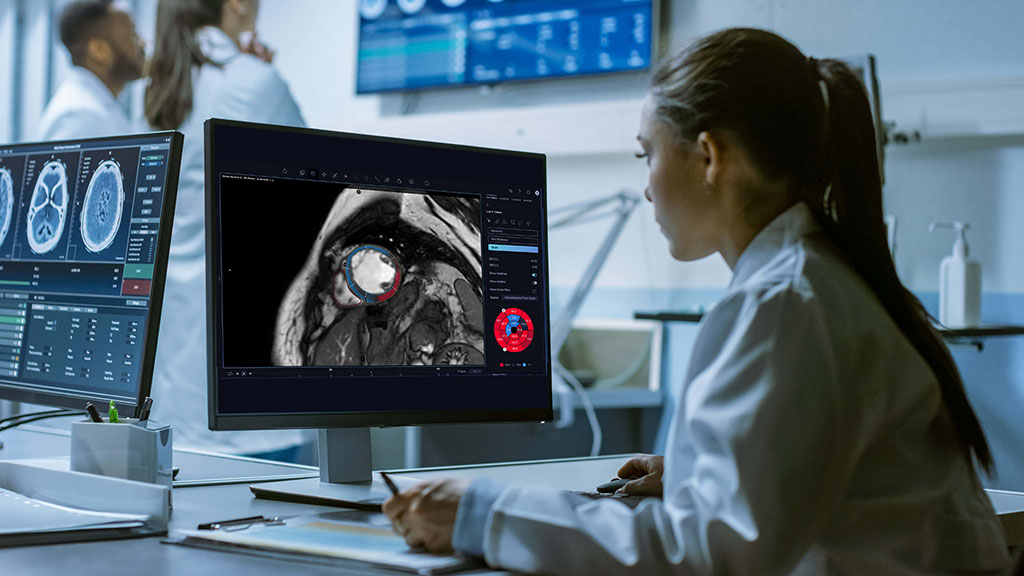
A comprehensive, cloud-based post-processing solution leverages AI and deep learning to provide extremely accurate, automated, fast, repeatable analysis of cardiac MRI images that are as accurate as segmentations performed manually by expert physicians.
Arterys’ (San Francisco, CA, USA) Cardio AI quantifies and visualizes 4D flow in minutes, enabling physicians to decrease scan time by ~30% and to maintain reimbursements, thus increasing the cost-effectiveness and ROI. By incorporating AI across the cardiac MR workflow, Cardio AI provides physicians with fast access to automated and quantitative cardiac MR image analysis and customizable reporting that reduces tedious and operator-dependent manual tasks, and saves up to 25 minutes per study.
Through the use of deep learning, Cardio AI provides automated, editable ventricular segmentations based on cine cardiac MRI images that are as accurate as segmentations performed manually by expert physicians. Cardio AI does not require on-premises hardware and results are accessible from anywhere and any device through a single user-interface zero footprint viewer and an internet connection. It seamlessly integrates with Powerscribe or Fluency dictation software ensuring exams are read accordingly and consistently.
Arterys has now announced several new modules to its Cardio AI clinical application and an additional FDA AI clearance based on deep learning. The latest Cardio AI enhancements provide physicians with additional tools to remove redundant work and tedious editing to analyze Cardiac MRI images. These new modules make it easier for physicians to manage the increasing demand and provide consistent diagnostic decisions more efficiently.
For instance, the new Cardio AI premium feature, Strain + AIPowered by 4D Flow provides cardiologists and radiologists with fast and reliable automated myocardial strain analysis. Another new feature allows cardiologists and radiologists to easily quantify volumes for both left and right atria in cardiac MRI images to aid in the diagnostic process. Arterys' Cardio AI clinical application has added another new feature that provides automatic wall thickness measurements to aid in the diagnosis process.
In addition to the Cardio AI enhancements, Arterys received its eighth FDA clearance. This additional FDA clearance makes Cardio AI's existing T1 + AI, and T2 quantification modules available for commercial use. These now-cleared modules offer a dedicated workflow for T1 and ECV, and T2 quantification, including multiple and global ROIs, and a graphical display of curves that provides users with intuitive, simple calculations for tissue characterization.
"Cardio AI is an integral part of our image interpretation and patient management," said Ali B Syed, M.D., at Stanford School of Medicine, Department of Radiology. "It significantly reduces the time necessary to generate useful information from our imaging. What used to take 30 minutes or more is now done, in many cases, before the patient is even off of the MRI table. It has also accelerated our adoption of techniques like T1 mapping and quantitative delayed enhancement because it dramatically reduces the effort to report these values."
"Cardio AI has been an invaluable tool in our practice to allow us faster processing of complex cardiac MR examinations with automated contouring not only for function but also tissue characterization saving us minutes of processing time on every single exam," said Melany B. Atkins, M.D., Division Chief, Body/Cardiovascular Imaging at Inova Fairfax Hospital. "Rapid processing of 4D Flow affords us the ability to evaluate complex congenital heart disease nearly real-time."
"With the addition of our new modules, we are able to accommodate a wide variety of users from community hospitals/physician centers to larger research centers and even pediatrics," said Michelle Buekes, Product Manager, Cardio AI at Arterys. "We are very excited to continue to work with our customers and partners to build upon and improve our product to meet the needs of an evolving and growing field."
Related Links:
Arterys
Latest MRI News
- New MRI Technique Reveals True Heart Age to Prevent Attacks and Strokes
- AI Tool Predicts Relapse of Pediatric Brain Cancer from Brain MRI Scans
- AI Tool Tracks Effectiveness of Multiple Sclerosis Treatments Using Brain MRI Scans
- Ultra-Powerful MRI Scans Enable Life-Changing Surgery in Treatment-Resistant Epileptic Patients
- AI-Powered MRI Technology Improves Parkinson’s Diagnoses
- Biparametric MRI Combined with AI Enhances Detection of Clinically Significant Prostate Cancer
- First-Of-Its-Kind AI-Driven Brain Imaging Platform to Better Guide Stroke Treatment Options
- New Model Improves Comparison of MRIs Taken at Different Institutions
- Groundbreaking New Scanner Sees 'Previously Undetectable' Cancer Spread
- First-Of-Its-Kind Tool Analyzes MRI Scans to Measure Brain Aging
- AI-Enhanced MRI Images Make Cancerous Breast Tissue Glow
- AI Model Automatically Segments MRI Images
- New Research Supports Routine Brain MRI Screening in Asymptomatic Late-Stage Breast Cancer Patients
- Revolutionary Portable Device Performs Rapid MRI-Based Stroke Imaging at Patient's Bedside
- AI Predicts After-Effects of Brain Tumor Surgery from MRI Scans
- MRI-First Strategy for Prostate Cancer Detection Proven Safe
Channels
Radiography
view channel
AI Improves Early Detection of Interval Breast Cancers
Interval breast cancers, which occur between routine screenings, are easier to treat when detected earlier. Early detection can reduce the need for aggressive treatments and improve the chances of better outcomes.... Read more
World's Largest Class Single Crystal Diamond Radiation Detector Opens New Possibilities for Diagnostic Imaging
Diamonds possess ideal physical properties for radiation detection, such as exceptional thermal and chemical stability along with a quick response time. Made of carbon with an atomic number of six, diamonds... Read moreUltrasound
view channel.jpeg)
AI-Powered Lung Ultrasound Outperforms Human Experts in Tuberculosis Diagnosis
Despite global declines in tuberculosis (TB) rates in previous years, the incidence of TB rose by 4.6% from 2020 to 2023. Early screening and rapid diagnosis are essential elements of the World Health... Read more
AI Identifies Heart Valve Disease from Common Imaging Test
Tricuspid regurgitation is a condition where the heart's tricuspid valve does not close completely during contraction, leading to backward blood flow, which can result in heart failure. A new artificial... Read moreNuclear Medicine
view channel
Novel Radiolabeled Antibody Improves Diagnosis and Treatment of Solid Tumors
Interleukin-13 receptor α-2 (IL13Rα2) is a cell surface receptor commonly found in solid tumors such as glioblastoma, melanoma, and breast cancer. It is minimally expressed in normal tissues, making it... Read more
Novel PET Imaging Approach Offers Never-Before-Seen View of Neuroinflammation
COX-2, an enzyme that plays a key role in brain inflammation, can be significantly upregulated by inflammatory stimuli and neuroexcitation. Researchers suggest that COX-2 density in the brain could serve... Read moreGeneral/Advanced Imaging
view channel
CT-Based Deep Learning-Driven Tool to Enhance Liver Cancer Diagnosis
Medical imaging, such as computed tomography (CT) scans, plays a crucial role in oncology, offering essential data for cancer detection, treatment planning, and monitoring of response to therapies.... Read more
AI-Powered Imaging System Improves Lung Cancer Diagnosis
Given the need to detect lung cancer at earlier stages, there is an increasing need for a definitive diagnostic pathway for patients with suspicious pulmonary nodules. However, obtaining tissue samples... Read moreImaging IT
view channel
New Google Cloud Medical Imaging Suite Makes Imaging Healthcare Data More Accessible
Medical imaging is a critical tool used to diagnose patients, and there are billions of medical images scanned globally each year. Imaging data accounts for about 90% of all healthcare data1 and, until... Read more
Global AI in Medical Diagnostics Market to Be Driven by Demand for Image Recognition in Radiology
The global artificial intelligence (AI) in medical diagnostics market is expanding with early disease detection being one of its key applications and image recognition becoming a compelling consumer proposition... Read moreIndustry News
view channel
GE HealthCare and NVIDIA Collaboration to Reimagine Diagnostic Imaging
GE HealthCare (Chicago, IL, USA) has entered into a collaboration with NVIDIA (Santa Clara, CA, USA), expanding the existing relationship between the two companies to focus on pioneering innovation in... Read more
Patient-Specific 3D-Printed Phantoms Transform CT Imaging
New research has highlighted how anatomically precise, patient-specific 3D-printed phantoms are proving to be scalable, cost-effective, and efficient tools in the development of new CT scan algorithms... Read more
Siemens and Sectra Collaborate on Enhancing Radiology Workflows
Siemens Healthineers (Forchheim, Germany) and Sectra (Linköping, Sweden) have entered into a collaboration aimed at enhancing radiologists' diagnostic capabilities and, in turn, improving patient care... Read more


