High Performance MRI Enhances Patient Experience
|
By MedImaging International staff writers Posted on 15 Dec 2021 |

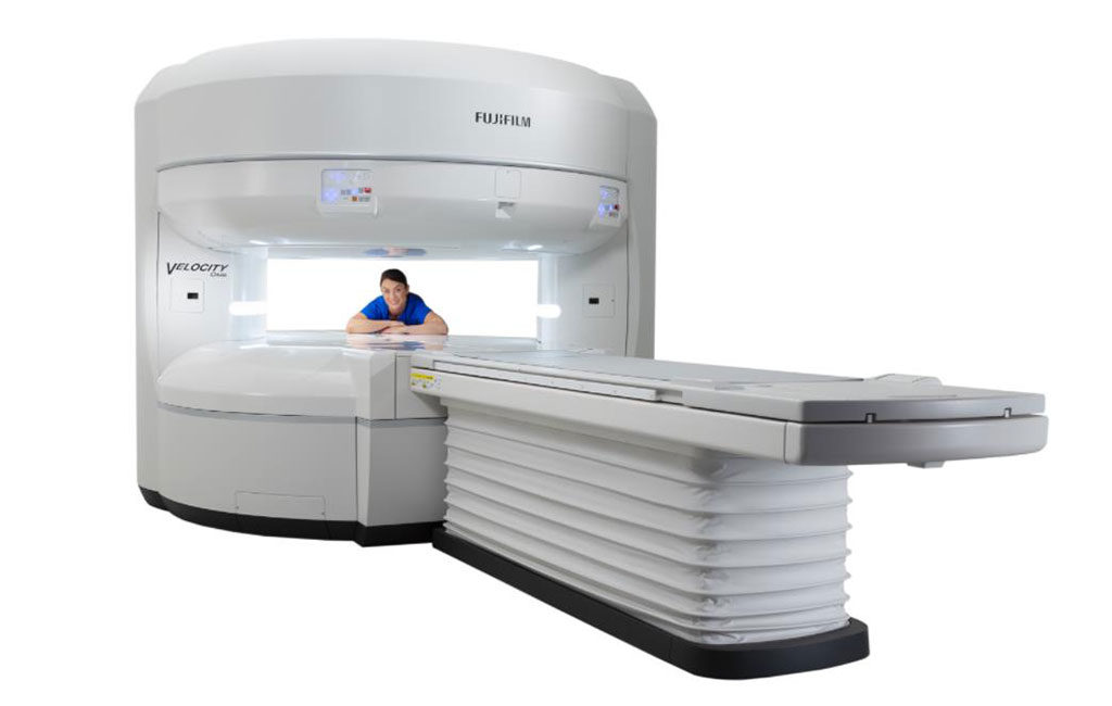
Image: The new Velocity 1.5 Tesla open MRI scanner (Photo courtesy of FujiFilm)
A brand new magnetic resonance imaging (MRI) system couples the advantages of a high-field scanner and the comfort of open MRI.
The Fujifilm Medical Systems (Tokyo, Japan) Velocity MRI is a 1.5 Tesla scanner that offers an open gantry design to create a feeling of spaciousness for bariatric, claustrophobic, geriatric, and pediatric patients; the open gantry also provides easy access for real-time interventional procedures. The extra-wide patient table can accommodate patients up to 300 kg, with in-gantry left and right movement and multiple coil connectors for easy positioning. The coils do not need to be swapped out, and can be left on the table between patients.
The symmetrical design of the system also enables it to be easily rotated with respect to the patient table. This creates an open lateral setting, which is ideal for both general and orthopedic imaging, as can be used to position patients comfortably, with their anatomy at the center of the magnet. Doing so helps to improve overall image quality, as the patient is able to lie comfortably and does not have to readjust their positon to get a good scan, as they might have to in a conventional bore system.
The Velocity MRI System also comes with integrated radiofrequency (RF) coils in order to streamline workflow and improve the patient experience, including the anatomy-conformable Synergy Flex coil for abdominal and orthopedic imaging in an open, vertical-field MRI scanner; it also has blanket-type RF coils for abdominal imaging and scanning of the knees and hips. Included is proprietary IP-RAPID iterative reconstruction technology, which accelerates 2D scans substantially and enables exams to be completed in 25%, 35%, or 40% of the current length of time.
“Velocity defines what today’s true Open MRI is about - unique patient comfort and accommodation benefits of open-sided MRI, now joined by the workflow and image quality benefits of integrated RF coils and enhanced reconstruction technologies,” said Shawn Etheridge, director of CT and MRI marketing for Fujifilm Healthcare Americas. “We now offer a scanner that can differentiate a hospital or outpatient imaging service, while delivering outstanding image quality, short exam times and operator ease-of-use benefits.”
Related Links:
Fujifilm Medical Systems
Latest MRI News
- New MRI Technique Reveals True Heart Age to Prevent Attacks and Strokes
- AI Tool Predicts Relapse of Pediatric Brain Cancer from Brain MRI Scans
- AI Tool Tracks Effectiveness of Multiple Sclerosis Treatments Using Brain MRI Scans
- Ultra-Powerful MRI Scans Enable Life-Changing Surgery in Treatment-Resistant Epileptic Patients
- AI-Powered MRI Technology Improves Parkinson’s Diagnoses
- Biparametric MRI Combined with AI Enhances Detection of Clinically Significant Prostate Cancer
- First-Of-Its-Kind AI-Driven Brain Imaging Platform to Better Guide Stroke Treatment Options
- New Model Improves Comparison of MRIs Taken at Different Institutions
- Groundbreaking New Scanner Sees 'Previously Undetectable' Cancer Spread
- First-Of-Its-Kind Tool Analyzes MRI Scans to Measure Brain Aging
- AI-Enhanced MRI Images Make Cancerous Breast Tissue Glow
- AI Model Automatically Segments MRI Images
- New Research Supports Routine Brain MRI Screening in Asymptomatic Late-Stage Breast Cancer Patients
- Revolutionary Portable Device Performs Rapid MRI-Based Stroke Imaging at Patient's Bedside
- AI Predicts After-Effects of Brain Tumor Surgery from MRI Scans
- MRI-First Strategy for Prostate Cancer Detection Proven Safe
Channels
Radiography
view channel
AI Improves Early Detection of Interval Breast Cancers
Interval breast cancers, which occur between routine screenings, are easier to treat when detected earlier. Early detection can reduce the need for aggressive treatments and improve the chances of better outcomes.... Read more
World's Largest Class Single Crystal Diamond Radiation Detector Opens New Possibilities for Diagnostic Imaging
Diamonds possess ideal physical properties for radiation detection, such as exceptional thermal and chemical stability along with a quick response time. Made of carbon with an atomic number of six, diamonds... Read moreUltrasound
view channel.jpeg)
AI-Powered Lung Ultrasound Outperforms Human Experts in Tuberculosis Diagnosis
Despite global declines in tuberculosis (TB) rates in previous years, the incidence of TB rose by 4.6% from 2020 to 2023. Early screening and rapid diagnosis are essential elements of the World Health... Read more
AI Identifies Heart Valve Disease from Common Imaging Test
Tricuspid regurgitation is a condition where the heart's tricuspid valve does not close completely during contraction, leading to backward blood flow, which can result in heart failure. A new artificial... Read moreNuclear Medicine
view channel
Novel Radiolabeled Antibody Improves Diagnosis and Treatment of Solid Tumors
Interleukin-13 receptor α-2 (IL13Rα2) is a cell surface receptor commonly found in solid tumors such as glioblastoma, melanoma, and breast cancer. It is minimally expressed in normal tissues, making it... Read more
Novel PET Imaging Approach Offers Never-Before-Seen View of Neuroinflammation
COX-2, an enzyme that plays a key role in brain inflammation, can be significantly upregulated by inflammatory stimuli and neuroexcitation. Researchers suggest that COX-2 density in the brain could serve... Read moreGeneral/Advanced Imaging
view channel
CT-Based Deep Learning-Driven Tool to Enhance Liver Cancer Diagnosis
Medical imaging, such as computed tomography (CT) scans, plays a crucial role in oncology, offering essential data for cancer detection, treatment planning, and monitoring of response to therapies.... Read more
AI-Powered Imaging System Improves Lung Cancer Diagnosis
Given the need to detect lung cancer at earlier stages, there is an increasing need for a definitive diagnostic pathway for patients with suspicious pulmonary nodules. However, obtaining tissue samples... Read moreImaging IT
view channel
New Google Cloud Medical Imaging Suite Makes Imaging Healthcare Data More Accessible
Medical imaging is a critical tool used to diagnose patients, and there are billions of medical images scanned globally each year. Imaging data accounts for about 90% of all healthcare data1 and, until... Read more
Global AI in Medical Diagnostics Market to Be Driven by Demand for Image Recognition in Radiology
The global artificial intelligence (AI) in medical diagnostics market is expanding with early disease detection being one of its key applications and image recognition becoming a compelling consumer proposition... Read moreIndustry News
view channel
GE HealthCare and NVIDIA Collaboration to Reimagine Diagnostic Imaging
GE HealthCare (Chicago, IL, USA) has entered into a collaboration with NVIDIA (Santa Clara, CA, USA), expanding the existing relationship between the two companies to focus on pioneering innovation in... Read more
Patient-Specific 3D-Printed Phantoms Transform CT Imaging
New research has highlighted how anatomically precise, patient-specific 3D-printed phantoms are proving to be scalable, cost-effective, and efficient tools in the development of new CT scan algorithms... Read more
Siemens and Sectra Collaborate on Enhancing Radiology Workflows
Siemens Healthineers (Forchheim, Germany) and Sectra (Linköping, Sweden) have entered into a collaboration aimed at enhancing radiologists' diagnostic capabilities and, in turn, improving patient care... Read more


