RSNA Officially Publishes First Dataset of Annotated COVID-19 Images
|
By MedImaging International staff writers Posted on 22 Dec 2020 |

Illustration
The Radiological Society of North America (RSNA; Oak Brook, IL, USA) and the RSNA COVID-19 AI Task Force have announced that the first annotated data set from the RSNA International COVID-19 Open Radiology Database (RICORD) has been published by The Cancer Imaging Archive (TCIA).
Radiologists have played a pivotal role in managing the pandemic, particularly when other testing methods are unavailable or when clinicians seek imaging data to inform treatment decisions. Although prediction models for COVID-19 imaging have been developed to support medical decision making, the lack of a diverse annotated data set has hindered the capabilities of these models. RSNA launched RICORD in mid-2020 with the goal of building the largest open database of anonymized COVID-19 medical images in the world. It is being made freely available to the global research and education communities to gain new insights, apply new tools such as artificial intelligence and deep learning, and accelerate clinical recognition of this novel disease.
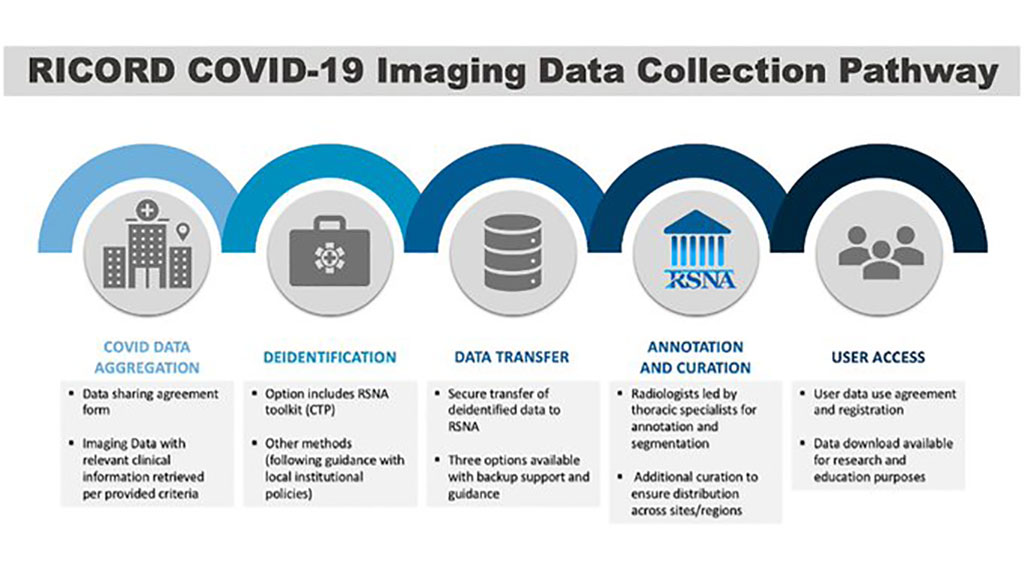
The RSNA COVID-19 AI Task Force hopes that RICORD will serve as a definitive source for COVID-19 imaging data by combining the contributions and experiences of medical imaging specialists and radiology departments worldwide. The RICORD data collection pathway enables radiology organizations to contribute data to RICORD safely and conveniently. It provides sites with guidance for data sharing and serves to standardize exam parameters, disease annotation terminology and clinical variables across these global efforts. Importantly, it connects to sustainable storage infrastructure via the US National Institutes of Health.
Created through a collaboration between RSNA and the Society of Thoracic Radiology, the initial group consists of 120 COVID-19 positive chest CT images from four international sites. This data set represents the first published component of RICORD, and RSNA’s first contribution to the Medical Imaging and Data Resource Center (MIDRC), a consortium for rapid and flexible collection, artificial intelligence analysis and dissemination of imaging and associated data. The RSNA COVID-19 AI Task Force will continue to update and expand both the volume and variety of data available in RICORD. A collection of COVID-19 negative chest CT control cases is in the pipeline for publication soon, along with a labeled set of 1,000 COVID-19 positive chest X-rays. An even larger set of CT and X-ray images has been submitted to RICORD and is currently being processed.
“RSNA was able to draw on relationships established from prior machine learning challenges to quickly put together a COVID-19 AI Task Force,” said Carol Wu, M.D., a radiologist at MD Anderson Cancer Center and a member of the RSNA task force. “Contributing sites, already proficient at sharing data with RSNA, were able to quickly process necessary legal agreements, identify suitable cases, perform image de-identification and transfer the images in record speed.”
“RSNA is extremely proud to be part of the MIDRC effort,” said Curtis Langlotz, M.D., Ph.D., RSNA Board liaison for information technology and annual meeting. “It will build a valuable repository of data for research to address the current pandemic and will serve as a model for how to collect and aggregate data to support imaging research.”
Related Links:
Radiological Society of North America
Latest Industry News News
- GE HealthCare and NVIDIA Collaboration to Reimagine Diagnostic Imaging
- Patient-Specific 3D-Printed Phantoms Transform CT Imaging
- Siemens and Sectra Collaborate on Enhancing Radiology Workflows
- Bracco Diagnostics and ColoWatch Partner to Expand Availability CRC Screening Tests Using Virtual Colonoscopy
- Mindray Partners with TeleRay to Streamline Ultrasound Delivery
- Philips and Medtronic Partner on Stroke Care
- Siemens and Medtronic Enter into Global Partnership for Advancing Spine Care Imaging Technologies
- RSNA 2024 Technical Exhibits to Showcase Latest Advances in Radiology
- Bracco Collaborates with Arrayus on Microbubble-Assisted Focused Ultrasound Therapy for Pancreatic Cancer
- Innovative Collaboration to Enhance Ischemic Stroke Detection and Elevate Standards in Diagnostic Imaging
- RSNA 2024 Registration Opens
- Microsoft collaborates with Leading Academic Medical Systems to Advance AI in Medical Imaging
- GE HealthCare Acquires Intelligent Ultrasound Group’s Clinical Artificial Intelligence Business
- Bayer and Rad AI Collaborate on Expanding Use of Cutting Edge AI Radiology Operational Solutions
- Polish Med-Tech Company BrainScan to Expand Extensively into Foreign Markets
- Hologic Acquires UK-Based Breast Surgical Guidance Company Endomagnetics Ltd.
Channels
Radiography
view channel
World's Largest Class Single Crystal Diamond Radiation Detector Opens New Possibilities for Diagnostic Imaging
Diamonds possess ideal physical properties for radiation detection, such as exceptional thermal and chemical stability along with a quick response time. Made of carbon with an atomic number of six, diamonds... Read more
AI-Powered Imaging Technique Shows Promise in Evaluating Patients for PCI
Percutaneous coronary intervention (PCI), also known as coronary angioplasty, is a minimally invasive procedure where small metal tubes called stents are inserted into partially blocked coronary arteries... Read moreMRI
view channel
AI Tool Predicts Relapse of Pediatric Brain Cancer from Brain MRI Scans
Many pediatric gliomas are treatable with surgery alone, but relapses can be catastrophic. Predicting which patients are at risk for recurrence remains challenging, leading to frequent follow-ups with... Read more
AI Tool Tracks Effectiveness of Multiple Sclerosis Treatments Using Brain MRI Scans
Multiple sclerosis (MS) is a condition in which the immune system attacks the brain and spinal cord, leading to impairments in movement, sensation, and cognition. Magnetic Resonance Imaging (MRI) markers... Read more
Ultra-Powerful MRI Scans Enable Life-Changing Surgery in Treatment-Resistant Epileptic Patients
Approximately 360,000 individuals in the UK suffer from focal epilepsy, a condition in which seizures spread from one part of the brain. Around a third of these patients experience persistent seizures... Read moreUltrasound
view channel.jpeg)
AI-Powered Lung Ultrasound Outperforms Human Experts in Tuberculosis Diagnosis
Despite global declines in tuberculosis (TB) rates in previous years, the incidence of TB rose by 4.6% from 2020 to 2023. Early screening and rapid diagnosis are essential elements of the World Health... Read more
AI Identifies Heart Valve Disease from Common Imaging Test
Tricuspid regurgitation is a condition where the heart's tricuspid valve does not close completely during contraction, leading to backward blood flow, which can result in heart failure. A new artificial... Read moreNuclear Medicine
view channel
Novel Radiolabeled Antibody Improves Diagnosis and Treatment of Solid Tumors
Interleukin-13 receptor α-2 (IL13Rα2) is a cell surface receptor commonly found in solid tumors such as glioblastoma, melanoma, and breast cancer. It is minimally expressed in normal tissues, making it... Read more
Novel PET Imaging Approach Offers Never-Before-Seen View of Neuroinflammation
COX-2, an enzyme that plays a key role in brain inflammation, can be significantly upregulated by inflammatory stimuli and neuroexcitation. Researchers suggest that COX-2 density in the brain could serve... Read moreGeneral/Advanced Imaging
view channel
AI-Powered Imaging System Improves Lung Cancer Diagnosis
Given the need to detect lung cancer at earlier stages, there is an increasing need for a definitive diagnostic pathway for patients with suspicious pulmonary nodules. However, obtaining tissue samples... Read more
AI Model Significantly Enhances Low-Dose CT Capabilities
Lung cancer remains one of the most challenging diseases, making early diagnosis vital for effective treatment. Fortunately, advancements in artificial intelligence (AI) are revolutionizing lung cancer... Read moreImaging IT
view channel
New Google Cloud Medical Imaging Suite Makes Imaging Healthcare Data More Accessible
Medical imaging is a critical tool used to diagnose patients, and there are billions of medical images scanned globally each year. Imaging data accounts for about 90% of all healthcare data1 and, until... Read more
Global AI in Medical Diagnostics Market to Be Driven by Demand for Image Recognition in Radiology
The global artificial intelligence (AI) in medical diagnostics market is expanding with early disease detection being one of its key applications and image recognition becoming a compelling consumer proposition... Read moreIndustry News
view channel
GE HealthCare and NVIDIA Collaboration to Reimagine Diagnostic Imaging
GE HealthCare (Chicago, IL, USA) has entered into a collaboration with NVIDIA (Santa Clara, CA, USA), expanding the existing relationship between the two companies to focus on pioneering innovation in... Read more
Patient-Specific 3D-Printed Phantoms Transform CT Imaging
New research has highlighted how anatomically precise, patient-specific 3D-printed phantoms are proving to be scalable, cost-effective, and efficient tools in the development of new CT scan algorithms... Read more
Siemens and Sectra Collaborate on Enhancing Radiology Workflows
Siemens Healthineers (Forchheim, Germany) and Sectra (Linköping, Sweden) have entered into a collaboration aimed at enhancing radiologists' diagnostic capabilities and, in turn, improving patient care... Read more


