Innovative RT System Could Treat Multiple Solid Tumors
|
By MedImaging International staff writers Posted on 20 Apr 2020 |
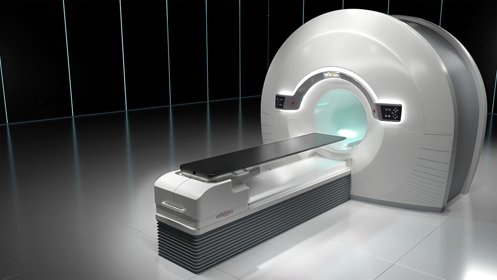
Image: The RefleXion Medical X1 machine (Photo courtesy of RefleXion Medical)
A groundbreaking radiotherapy (RT) device rotates up to 60 times faster than other linear accelerators and modulates dose delivery from 100 points per beam station.
The RefleXion Medical (Hayward, CA, USA) X1 machine is intended for image-guided RT, including stereotactic body radiotherapy (SBRT), stereotactic radiosurgery (SRS), intensity modulated radiotherapy (IMRT), and in the future, biology-guided radiotherapy (BgRT). The multiple indications are achieved by combining high quality fan-beam computed tomography (CT) imaging with a linear accelerator (LINAC) in order to reduce motion artifacts that can inhibit accurate targeting of the radiation dose to a patient’s tumor.
The hybrid imaging-therapy system can define field sizes based on jaws that are 40 cm wide, with slice width choices of either one or two cm to optimize patient treatment. A binary multileaf collimator (MLC) is used to create intensity modulated radiation fields at a source-axis distance of 85 cm. Clinical sub-systems include 6 MV photon RT delivery, Kilovoltage level X-ray CT imaging, MV X-ray detection, and treatment planning. The incorporated LINAC, along with a fixed primary collimator, several adjustable collimators, and the MLC are mounted in the therapy plane.
The system is also designed for inclusion of positron emission tomography (PET) imaging components to enable BgRT, which uses the biological signature of each tumor to characterize its movement and to track tumors more precisely. The RefleXion X1 machine with BgRT intends to overcome the technical limitations that currently restrict RT to one or two tumors. Once developed, RefleXion will scale BgRT on the X1 machine to treat all visible tumors, even those that move rapidly due to bodily functions such as breathing or digestion, in the same treatment session.
“The idea was to use individual emissions that make up a partially formed PET image, and are generated by the tumor, as a homing signal or biological marker to quickly guide radiotherapy to that location,” said Sam Mazin, PhD, co-founder and chief technology officer of RefleXion. “In essence, [it is] turning cancer on itself to destroy it. It has been a rewarding journey from thinking of the idea years ago to co-founding RefleXion.”
SRS is a non-surgical radiation therapy used to treat functional abnormalities and small tumors of the brain. When SRS is used to treat body tumors, it is called stereotactic body radiotherapy (SBRT). Both SRS and SBRT are an image-guided radiation therapy (IGRT), relying on 3D imaging and localization techniques, immobilization systems for precise patient position during therapy, and highly focused gamma-ray or x-ray beams that converge on the tumor.
Related Links:
RefleXion Medical
Latest Nuclear Medicine News
- Novel Bacteria-Specific PET Imaging Approach Detects Hard-To-Diagnose Lung Infections
- New Imaging Approach Could Reduce Need for Biopsies to Monitor Prostate Cancer
- Novel Radiolabeled Antibody Improves Diagnosis and Treatment of Solid Tumors
- Novel PET Imaging Approach Offers Never-Before-Seen View of Neuroinflammation
- Novel Radiotracer Identifies Biomarker for Triple-Negative Breast Cancer
- Innovative PET Imaging Technique to Help Diagnose Neurodegeneration
- New Molecular Imaging Test to Improve Lung Cancer Diagnosis
- Novel PET Technique Visualizes Spinal Cord Injuries to Predict Recovery
- Next-Gen Tau Radiotracers Outperform FDA-Approved Imaging Agents in Detecting Alzheimer’s
- Breakthrough Method Detects Inflammation in Body Using PET Imaging
- Advanced Imaging Reveals Hidden Metastases in High-Risk Prostate Cancer Patients
- Combining Advanced Imaging Technologies Offers Breakthrough in Glioblastoma Treatment
- New Molecular Imaging Agent Accurately Identifies Crucial Cancer Biomarker
- New Scans Light Up Aggressive Tumors for Better Treatment
- AI Stroke Brain Scan Readings Twice as Accurate as Current Method
- AI Analysis of PET/CT Images Predicts Side Effects of Immunotherapy in Lung Cancer
Channels
Radiography
view channel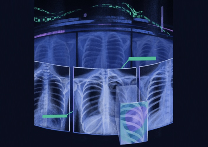
New AI Tool Helps Doctors Read Chest X‑Rays Better
The U.S. health care system ranks poorly in critical health indicators, including life expectancy, and patients often seek better outcomes with fewer costs. Doctors aim for precise, first-time diagnoses,... Read more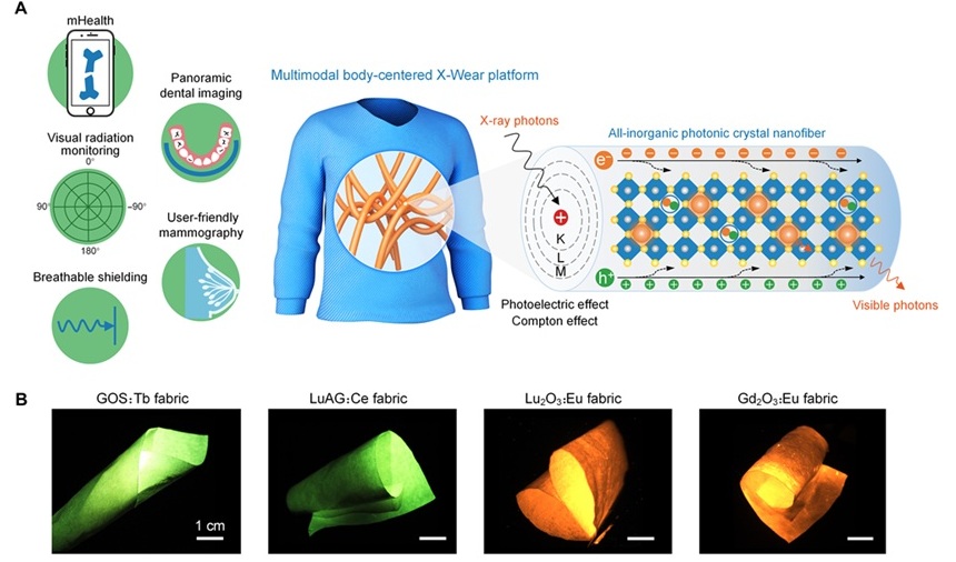
Wearable X-Ray Imaging Detecting Fabric to Provide On-The-Go Diagnostic Scanning
X-rays have been instrumental in modern medical diagnostics since their discovery, from imaging broken bones to screening for early signs of breast cancer. However, traditional X-ray detectors, primarily... Read moreMRI
view channel
AI-Assisted Model Enhances MRI Heart Scans
A cardiac MRI can reveal critical information about the heart’s function and any abnormalities, but traditional scans take 30 to 90 minutes and often suffer from poor image quality due to patient movement.... Read more
AI Model Outperforms Doctors at Identifying Patients Most At-Risk of Cardiac Arrest
Hypertrophic cardiomyopathy is one of the most common inherited heart conditions and a leading cause of sudden cardiac death in young individuals and athletes. While many patients live normal lives, some... Read moreUltrasound
view channel
Non-Invasive Ultrasound-Based Tool Accurately Detects Infant Meningitis
Meningitis, an inflammation of the membranes surrounding the brain and spinal cord, can be fatal in infants if not diagnosed and treated early. Even when treated, it may leave lasting damage, such as cognitive... Read more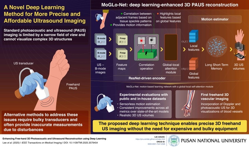
Breakthrough Deep Learning Model Enhances Handheld 3D Medical Imaging
Ultrasound imaging is a vital diagnostic technique used to visualize internal organs and tissues in real time and to guide procedures such as biopsies and injections. When paired with photoacoustic imaging... Read moreGeneral/Advanced Imaging
view channel
CT Colonography Beats Stool DNA Testing for Colon Cancer Screening
As colorectal cancer remains the second leading cause of cancer-related deaths worldwide, early detection through screening is vital to reduce advanced-stage treatments and associated costs.... Read more
First-Of-Its-Kind Wearable Device Offers Revolutionary Alternative to CT Scans
Currently, patients with conditions such as heart failure, pneumonia, or respiratory distress often require multiple imaging procedures that are intermittent, disruptive, and involve high levels of radiation.... Read more
AI-Based CT Scan Analysis Predicts Early-Stage Kidney Damage Due to Cancer Treatments
Radioligand therapy, a form of targeted nuclear medicine, has recently gained attention for its potential in treating specific types of tumors. However, one of the potential side effects of this therapy... Read moreImaging IT
view channel
New Google Cloud Medical Imaging Suite Makes Imaging Healthcare Data More Accessible
Medical imaging is a critical tool used to diagnose patients, and there are billions of medical images scanned globally each year. Imaging data accounts for about 90% of all healthcare data1 and, until... Read more
Global AI in Medical Diagnostics Market to Be Driven by Demand for Image Recognition in Radiology
The global artificial intelligence (AI) in medical diagnostics market is expanding with early disease detection being one of its key applications and image recognition becoming a compelling consumer proposition... Read moreIndustry News
view channel
GE HealthCare and NVIDIA Collaboration to Reimagine Diagnostic Imaging
GE HealthCare (Chicago, IL, USA) has entered into a collaboration with NVIDIA (Santa Clara, CA, USA), expanding the existing relationship between the two companies to focus on pioneering innovation in... Read more
Patient-Specific 3D-Printed Phantoms Transform CT Imaging
New research has highlighted how anatomically precise, patient-specific 3D-printed phantoms are proving to be scalable, cost-effective, and efficient tools in the development of new CT scan algorithms... Read more
Siemens and Sectra Collaborate on Enhancing Radiology Workflows
Siemens Healthineers (Forchheim, Germany) and Sectra (Linköping, Sweden) have entered into a collaboration aimed at enhancing radiologists' diagnostic capabilities and, in turn, improving patient care... Read more


