Optical Hydrogel Monitors Cancer Patients Radiation Dose
|
By MedImaging International staff writers Posted on 26 Feb 2020 |

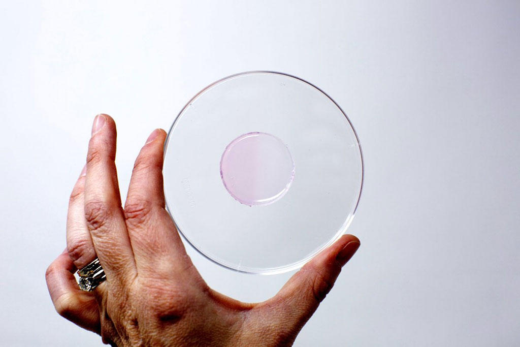
Image: A circle of the hydrogel, irradiated on the left half, whereas the right half is not irradiated (Photo courtesy of ASU)
A novel hydrogel applied directly to a patient's skin changes color in direct correlation to radiation therapy (RT) dose levels, claims a new study.
Developed by researchers at Arizona State University (ASU; Tempe, USA) and Banner-M.D. Anderson Cancer Center (Gilbert, AZ, USA), the gel-based nanosensor is impregnated with gold salts and amino acids. Exposure to ionizing radiation results in the conversion of gold ions in the gel to gold nanoparticles, which render a visual change in color in the gel due to their plasmonic properties. Without radiation, the hydrogel is colorless; but as it is exposed to radiation, it turns pink, with the color intensity directly correlated to the amount of radiation.
The gel nanosensor can detect complex topographical dose patterns, with the intensity of color formed in the gel serving as a quantitative reporter of the ionizing radiation. At the end of RT therapy, the gel is painlessly peeled off the skin and the color hue is measured with the aid of an absorption spectrometer. The gel has so far been tested on an anthropomorphic phantom and in live dogs undergoing clinical grade RT. The study was presented at the 64th annual meeting of the Biophysical Society, held during February 2020 in San Diego (CA, USA).
“The ease of fabrication, operation, rapid readout, colorimetric detection, and relatively low cost illustrate the translational potential of this technology for topographical dose mapping in radiotherapy applications in the clinic,” said lead author and study presenter Subhadeep Dutta, MSc, of ASU. “Our next plan is to convert it to an app-based system, where you can take a picture of a gel and that can predict the dose based on programming in the app. It's just measuring color, which is easy to do.”
Examples of current dose monitors include radiochromic films, which resemble a sheet of paper; but as they are sensitive to light and heat, they must be carefully handled, and require long processing times. Other methods include quantum dots and metal organic frameworks, which demonstrate an intense scintillating response, but provide only point dose information; and polymer gel dosimeters that rely on sophisticated readout techniques (such as MRI) for post-irradiation analysis.
Related Links:
Arizona State University
Banner-M.D. Anderson Cancer Center
Latest Nuclear Medicine News
- PET Imaging of Inflammation Predicts Recovery and Guides Therapy After Heart Attack
- Radiotheranostic Approach Detects, Kills and Reprograms Aggressive Cancers
- New Imaging Solution Improves Survival for Patients with Recurring Prostate Cancer
- PET Tracer Enables Same-Day Imaging of Triple-Negative Breast and Urothelial Cancers
- New Camera Sees Inside Human Body for Enhanced Scanning and Diagnosis
- Novel Bacteria-Specific PET Imaging Approach Detects Hard-To-Diagnose Lung Infections
- New Imaging Approach Could Reduce Need for Biopsies to Monitor Prostate Cancer
- Novel Radiolabeled Antibody Improves Diagnosis and Treatment of Solid Tumors
- Novel PET Imaging Approach Offers Never-Before-Seen View of Neuroinflammation
- Novel Radiotracer Identifies Biomarker for Triple-Negative Breast Cancer
- Innovative PET Imaging Technique to Help Diagnose Neurodegeneration
- New Molecular Imaging Test to Improve Lung Cancer Diagnosis
- Novel PET Technique Visualizes Spinal Cord Injuries to Predict Recovery
- Next-Gen Tau Radiotracers Outperform FDA-Approved Imaging Agents in Detecting Alzheimer’s
- Breakthrough Method Detects Inflammation in Body Using PET Imaging
- Advanced Imaging Reveals Hidden Metastases in High-Risk Prostate Cancer Patients
Channels
Radiography
view channel
X-Ray Breakthrough Captures Three Image-Contrast Types in Single Shot
Detecting early-stage cancer or subtle changes deep inside tissues has long challenged conventional X-ray systems, which rely only on how structures absorb radiation. This limitation keeps many microstructural... Read more
AI Generates Future Knee X-Rays to Predict Osteoarthritis Progression Risk
Osteoarthritis, a degenerative joint disease affecting over 500 million people worldwide, is the leading cause of disability among older adults. Current diagnostic tools allow doctors to assess damage... Read moreMRI
view channel
Novel Imaging Approach to Improve Treatment for Spinal Cord Injuries
Vascular dysfunction in the spinal cord contributes to multiple neurological conditions, including traumatic injuries and degenerative cervical myelopathy, where reduced blood flow can lead to progressive... Read more
AI-Assisted Model Enhances MRI Heart Scans
A cardiac MRI can reveal critical information about the heart’s function and any abnormalities, but traditional scans take 30 to 90 minutes and often suffer from poor image quality due to patient movement.... Read more
AI Model Outperforms Doctors at Identifying Patients Most At-Risk of Cardiac Arrest
Hypertrophic cardiomyopathy is one of the most common inherited heart conditions and a leading cause of sudden cardiac death in young individuals and athletes. While many patients live normal lives, some... Read moreUltrasound
view channel
Wearable Ultrasound Imaging System to Enable Real-Time Disease Monitoring
Chronic conditions such as hypertension and heart failure require close monitoring, yet today’s ultrasound imaging is largely confined to hospitals and short, episodic scans. This reactive model limits... Read more
Ultrasound Technique Visualizes Deep Blood Vessels in 3D Without Contrast Agents
Producing clear 3D images of deep blood vessels has long been difficult without relying on contrast agents, CT scans, or MRI. Standard ultrasound typically provides only 2D cross-sections, limiting clinicians’... Read moreGeneral/Advanced Imaging
view channel
3D Scanning Approach Enables Ultra-Precise Brain Surgery
Precise navigation is critical in neurosurgery, yet even small alignment errors can affect outcomes when operating deep within the brain. A new 3D surface-scanning approach now provides a radiation-free... Read more
AI Tool Improves Medical Imaging Process by 90%
Accurately labeling different regions within medical scans, a process known as medical image segmentation, is critical for diagnosis, surgery planning, and research. Traditionally, this has been a manual... Read more
New Ultrasmall, Light-Sensitive Nanoparticles Could Serve as Contrast Agents
Medical imaging technologies face ongoing challenges in capturing accurate, detailed views of internal processes, especially in conditions like cancer, where tracking disease development and treatment... Read more
AI Algorithm Accurately Predicts Pancreatic Cancer Metastasis Using Routine CT Images
In pancreatic cancer, detecting whether the disease has spread to other organs is critical for determining whether surgery is appropriate. If metastasis is present, surgery is not recommended, yet current... Read moreImaging IT
view channel
New Google Cloud Medical Imaging Suite Makes Imaging Healthcare Data More Accessible
Medical imaging is a critical tool used to diagnose patients, and there are billions of medical images scanned globally each year. Imaging data accounts for about 90% of all healthcare data1 and, until... Read more
Global AI in Medical Diagnostics Market to Be Driven by Demand for Image Recognition in Radiology
The global artificial intelligence (AI) in medical diagnostics market is expanding with early disease detection being one of its key applications and image recognition becoming a compelling consumer proposition... Read moreIndustry News
view channel
GE HealthCare and NVIDIA Collaboration to Reimagine Diagnostic Imaging
GE HealthCare (Chicago, IL, USA) has entered into a collaboration with NVIDIA (Santa Clara, CA, USA), expanding the existing relationship between the two companies to focus on pioneering innovation in... Read more
Patient-Specific 3D-Printed Phantoms Transform CT Imaging
New research has highlighted how anatomically precise, patient-specific 3D-printed phantoms are proving to be scalable, cost-effective, and efficient tools in the development of new CT scan algorithms... Read more
Siemens and Sectra Collaborate on Enhancing Radiology Workflows
Siemens Healthineers (Forchheim, Germany) and Sectra (Linköping, Sweden) have entered into a collaboration aimed at enhancing radiologists' diagnostic capabilities and, in turn, improving patient care... Read more


