AI Module Delivers Predictive Image Segmentation and Processing
|
By MedImaging International staff writers Posted on 23 Dec 2019 |

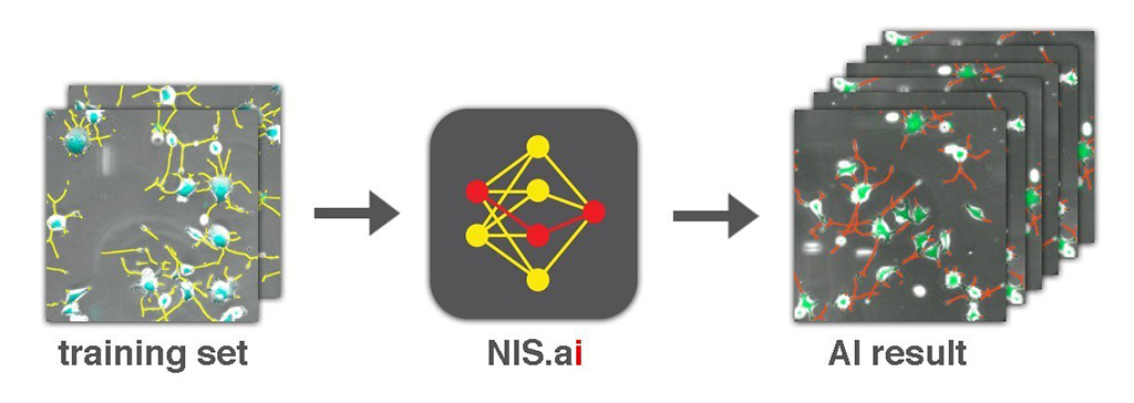
Image: A suite of microscopy applications aid predictive imaging, segmentation, and processing (Photo courtesy of Nikon Instruments)
A powerful image analysis and processing module leverages deep learning and artificial intelligence (AI) to accurately extract unbiased data from vast amounts of microscopy datasets.
The Nikon Instruments (Melville, NY, USA) NIS.ai microscopy image analysis and processing module is a suite of AI-based processing tools that utilizes convolutional neural networks (CNNs) in order to learn how to read images from small training datasets supplied by the user. The training results can then be applied to process and analyze huge volumes of data, allowing researchers to increase throughput and expand their application limits. The NIS.ai includes a suite of applications for predictive imaging, image segmentation, and processing. These include:
Convert.ai, which learns related patterns in two different imaging channels. After training, Convert.ai can predict the pattern in the second channel, even when presented with only the first channel. It can also be trained to predict where DAPI-based fluorescent staining of nuclei--a common method for cell segmentation and counting--could be based on unstained differential interference contrast (DIC) or phase-contrast microscopy images. This enables users to perform nuclei-based image analysis without ever having to stain samples with DAPI or acquire a fluorescent channel.
Segment.ai, which enables complex structures to be easily identified and segmented. Neurites in phase-contrast images are traditionally difficult to define by classic thresholding. Segment.ai can be trained on a small subset of hand-traced neurites to automatically detect and segment neurites from thousands of untraced datasets.
Enhance.ai, which allows dim fluorescent samples with poor signal-to-noise ratio (SNR) to be enhanced by learning what a high signal-to-noise image looks like, via a process that compares under-exposed and optimally-exposed images. Enhance.ai can then restore details in under-exposed or dim fluorescent images, enabling researchers to gain more insights from their low-signal imaging applications.
Denoise.ai, which removes shot noise from resonant confocal images and can be performed in real-time. Applying Denoise.ai to resonant confocal imaging enables users to acquire confocal images at ultra-high speed without sacrificing image quality.
“The application of Deep Learning and AI to biomedical imaging is extremely powerful, and opening up unseen possibilities,” said Steve Ross, PhD, director of products and marketing at Nikon Instruments. “With NIS.ai, researchers can easily apply deep learning to extract meaningful, unbiased data from large, complex datasets.”
Related Links:
Nikon Instruments
Latest General/Advanced Imaging News
- AI-Powered Imaging System Improves Lung Cancer Diagnosis
- AI Model Significantly Enhances Low-Dose CT Capabilities
- Ultra-Low Dose CT Aids Pneumonia Diagnosis in Immunocompromised Patients
- AI Reduces CT Lung Cancer Screening Workload by Almost 80%
- Cutting-Edge Technology Combines Light and Sound for Real-Time Stroke Monitoring
- AI System Detects Subtle Changes in Series of Medical Images Over Time
- New CT Scan Technique to Improve Prognosis and Treatments for Head and Neck Cancers
- World’s First Mobile Whole-Body CT Scanner to Provide Diagnostics at POC
- Comprehensive CT Scans Could Identify Atherosclerosis Among Lung Cancer Patients
- AI Improves Detection of Colorectal Cancer on Routine Abdominopelvic CT Scans
- Super-Resolution Technology Enhances Clinical Bone Imaging to Predict Osteoporotic Fracture Risk
- AI-Powered Abdomen Map Enables Early Cancer Detection
- Deep Learning Model Detects Lung Tumors on CT
- AI Predicts Cardiovascular Risk from CT Scans
- Deep Learning Based Algorithms Improve Tumor Detection in PET/CT Scans
- New Technology Provides Coronary Artery Calcification Scoring on Ungated Chest CT Scans
Channels
Radiography
view channel
World's Largest Class Single Crystal Diamond Radiation Detector Opens New Possibilities for Diagnostic Imaging
Diamonds possess ideal physical properties for radiation detection, such as exceptional thermal and chemical stability along with a quick response time. Made of carbon with an atomic number of six, diamonds... Read more
AI-Powered Imaging Technique Shows Promise in Evaluating Patients for PCI
Percutaneous coronary intervention (PCI), also known as coronary angioplasty, is a minimally invasive procedure where small metal tubes called stents are inserted into partially blocked coronary arteries... Read moreMRI
view channel
AI Tool Tracks Effectiveness of Multiple Sclerosis Treatments Using Brain MRI Scans
Multiple sclerosis (MS) is a condition in which the immune system attacks the brain and spinal cord, leading to impairments in movement, sensation, and cognition. Magnetic Resonance Imaging (MRI) markers... Read more
Ultra-Powerful MRI Scans Enable Life-Changing Surgery in Treatment-Resistant Epileptic Patients
Approximately 360,000 individuals in the UK suffer from focal epilepsy, a condition in which seizures spread from one part of the brain. Around a third of these patients experience persistent seizures... Read more
AI-Powered MRI Technology Improves Parkinson’s Diagnoses
Current research shows that the accuracy of diagnosing Parkinson’s disease typically ranges from 55% to 78% within the first five years of assessment. This is partly due to the similarities shared by Parkinson’s... Read more
Biparametric MRI Combined with AI Enhances Detection of Clinically Significant Prostate Cancer
Artificial intelligence (AI) technologies are transforming the way medical images are analyzed, offering unprecedented capabilities in quantitatively extracting features that go beyond traditional visual... Read moreUltrasound
view channel.jpeg)
AI-Powered Lung Ultrasound Outperforms Human Experts in Tuberculosis Diagnosis
Despite global declines in tuberculosis (TB) rates in previous years, the incidence of TB rose by 4.6% from 2020 to 2023. Early screening and rapid diagnosis are essential elements of the World Health... Read more
AI Identifies Heart Valve Disease from Common Imaging Test
Tricuspid regurgitation is a condition where the heart's tricuspid valve does not close completely during contraction, leading to backward blood flow, which can result in heart failure. A new artificial... Read moreNuclear Medicine
view channel
Novel PET Imaging Approach Offers Never-Before-Seen View of Neuroinflammation
COX-2, an enzyme that plays a key role in brain inflammation, can be significantly upregulated by inflammatory stimuli and neuroexcitation. Researchers suggest that COX-2 density in the brain could serve... Read more
Novel Radiotracer Identifies Biomarker for Triple-Negative Breast Cancer
Triple-negative breast cancer (TNBC), which represents 15-20% of all breast cancer cases, is one of the most aggressive subtypes, with a five-year survival rate of about 40%. Due to its significant heterogeneity... Read moreImaging IT
view channel
New Google Cloud Medical Imaging Suite Makes Imaging Healthcare Data More Accessible
Medical imaging is a critical tool used to diagnose patients, and there are billions of medical images scanned globally each year. Imaging data accounts for about 90% of all healthcare data1 and, until... Read more
Global AI in Medical Diagnostics Market to Be Driven by Demand for Image Recognition in Radiology
The global artificial intelligence (AI) in medical diagnostics market is expanding with early disease detection being one of its key applications and image recognition becoming a compelling consumer proposition... Read moreIndustry News
view channel
GE HealthCare and NVIDIA Collaboration to Reimagine Diagnostic Imaging
GE HealthCare (Chicago, IL, USA) has entered into a collaboration with NVIDIA (Santa Clara, CA, USA), expanding the existing relationship between the two companies to focus on pioneering innovation in... Read more
Patient-Specific 3D-Printed Phantoms Transform CT Imaging
New research has highlighted how anatomically precise, patient-specific 3D-printed phantoms are proving to be scalable, cost-effective, and efficient tools in the development of new CT scan algorithms... Read more
Siemens and Sectra Collaborate on Enhancing Radiology Workflows
Siemens Healthineers (Forchheim, Germany) and Sectra (Linköping, Sweden) have entered into a collaboration aimed at enhancing radiologists' diagnostic capabilities and, in turn, improving patient care... Read more


