New All-Optical System Could Revolutionize Image-Guided Interventions
|
By MedImaging International staff writers Posted on 29 Aug 2018 |

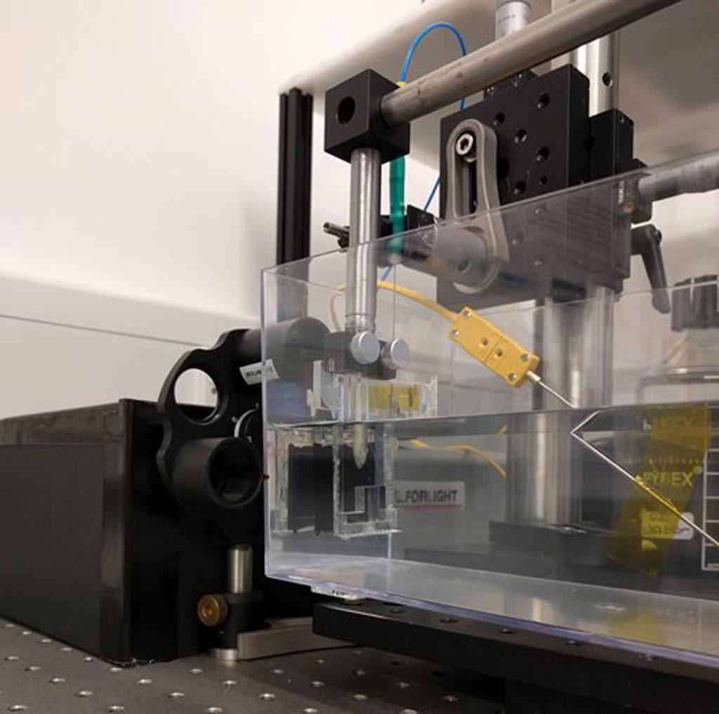
Image: All-optical ultrasound imager. This prototype is the first all-optical ultrasound imager to demonstrate video-rate, real-time 2D imaging of biological tissue. The achievement is an important step toward making all-optical ultrasound practical for routine clinical use, where it could offer significant advantages over current imaging technology (Photo courtesy of Erwin J. Alles, University College London).
A new ultrasound system that uses optical, instead of electronic components, could improve performance while providing doctors with significantly more flexibility in the way they use ultrasound to diagnose and treat medical problems. For the first time, researchers have demonstrated the use of an all-optical ultrasound imager for video-rate, real-time 2D imaging of biological tissue, marking a significant step towards making all-optical ultrasound practical for routine clinical use. Since all-optical ultrasound systems do not require any electronic components in the imaging probe, they could be safely used at the same time as magnetic resonance imaging (MRI) scanners. This can provide doctors with a more comprehensive picture of the tissues around an area of interest, such as a tumor or blood vessel, according to a recent paper published in the Optical Society (OSA) journal Biomedical Optics Express.
Conventional ultrasound imagers use arrays of electronic transducers to transmit high-frequency sound waves into tissue and receive the reflections, whereas all-optical ultrasound imagers use light to both transmit and receive ultrasound waves. Pulsed laser light is used to generate ultrasound waves, and scanning mirrors control where the waves are transmitted into the tissue. A fiber optic sensor then receives the reflected waves.
The researchers demonstrated how the light source can be manipulated to generate either low frequency ultrasound, resulting in greater penetration into the tissue, or high frequency ultrasound, which offers higher resolution images at a shallower depth. The team tested their prototype system by imaging a deceased zebrafish and a pig artery that was manipulated to emulate the dynamics of pulsing blood. The demonstration showed imaging capabilities comparable to an electronic high-frequency ultrasound system, with a sustained frame rate of 15 Hertz, a dynamic range of 30 decibels, a penetration depth of 6 millimeters and a resolution of 75 by 100 micrometers.
The researchers are now working to adapt the technology for clinical use by developing a long, flexible imaging probe for free-hand operation, as well as miniaturized versions for endoscopic applications. In conventional ultrasound devices, the electronic components make it difficult to miniaturize the devices for internal use. On the other hand, optical components are easily miniaturized and tiny all-optical ultrasound probes are likely be significantly less expensive to manufacture as compared to compact electronic ultrasound systems, according to the researchers.
“All-optical ultrasound imaging probes have the potential to revolutionize image-guided interventions,” said Erwin J. Alles, University College London. “A lack of electronics and the resulting MRI compatibility will allow for true multimodality image guidance, with probes that are potentially just a fraction of the cost of conventional electronic counterparts.”
Latest Industry News News
- GE HealthCare and NVIDIA Collaboration to Reimagine Diagnostic Imaging
- Patient-Specific 3D-Printed Phantoms Transform CT Imaging
- Siemens and Sectra Collaborate on Enhancing Radiology Workflows
- Bracco Diagnostics and ColoWatch Partner to Expand Availability CRC Screening Tests Using Virtual Colonoscopy
- Mindray Partners with TeleRay to Streamline Ultrasound Delivery
- Philips and Medtronic Partner on Stroke Care
- Siemens and Medtronic Enter into Global Partnership for Advancing Spine Care Imaging Technologies
- RSNA 2024 Technical Exhibits to Showcase Latest Advances in Radiology
- Bracco Collaborates with Arrayus on Microbubble-Assisted Focused Ultrasound Therapy for Pancreatic Cancer
- Innovative Collaboration to Enhance Ischemic Stroke Detection and Elevate Standards in Diagnostic Imaging
- RSNA 2024 Registration Opens
- Microsoft collaborates with Leading Academic Medical Systems to Advance AI in Medical Imaging
- GE HealthCare Acquires Intelligent Ultrasound Group’s Clinical Artificial Intelligence Business
- Bayer and Rad AI Collaborate on Expanding Use of Cutting Edge AI Radiology Operational Solutions
- Polish Med-Tech Company BrainScan to Expand Extensively into Foreign Markets
- Hologic Acquires UK-Based Breast Surgical Guidance Company Endomagnetics Ltd.
Channels
Radiography
view channel
AI-Powered Imaging Technique Shows Promise in Evaluating Patients for PCI
Percutaneous coronary intervention (PCI), also known as coronary angioplasty, is a minimally invasive procedure where small metal tubes called stents are inserted into partially blocked coronary arteries... Read more
Higher Chest X-Ray Usage Catches Lung Cancer Earlier and Improves Survival
Lung cancer continues to be the leading cause of cancer-related deaths worldwide. While advanced technologies like CT scanners play a crucial role in detecting lung cancer, more accessible and affordable... Read moreMRI
view channel
Ultra-Powerful MRI Scans Enable Life-Changing Surgery in Treatment-Resistant Epileptic Patients
Approximately 360,000 individuals in the UK suffer from focal epilepsy, a condition in which seizures spread from one part of the brain. Around a third of these patients experience persistent seizures... Read more
AI-Powered MRI Technology Improves Parkinson’s Diagnoses
Current research shows that the accuracy of diagnosing Parkinson’s disease typically ranges from 55% to 78% within the first five years of assessment. This is partly due to the similarities shared by Parkinson’s... Read more
Biparametric MRI Combined with AI Enhances Detection of Clinically Significant Prostate Cancer
Artificial intelligence (AI) technologies are transforming the way medical images are analyzed, offering unprecedented capabilities in quantitatively extracting features that go beyond traditional visual... Read more
First-Of-Its-Kind AI-Driven Brain Imaging Platform to Better Guide Stroke Treatment Options
Each year, approximately 800,000 people in the U.S. experience strokes, with marginalized and minoritized groups being disproportionately affected. Strokes vary in terms of size and location within the... Read moreUltrasound
view channel
Smart Ultrasound-Activated Immune Cells Destroy Cancer Cells for Extended Periods
Chimeric antigen receptor (CAR) T-cell therapy has emerged as a highly promising cancer treatment, especially for bloodborne cancers like leukemia. This highly personalized therapy involves extracting... Read more
Tiny Magnetic Robot Takes 3D Scans from Deep Within Body
Colorectal cancer ranks as one of the leading causes of cancer-related mortality worldwide. However, when detected early, it is highly treatable. Now, a new minimally invasive technique could significantly... Read more
High Resolution Ultrasound Speeds Up Prostate Cancer Diagnosis
Each year, approximately one million prostate cancer biopsies are conducted across Europe, with similar numbers in the USA and around 100,000 in Canada. Most of these biopsies are performed using MRI images... Read more
World's First Wireless, Handheld, Whole-Body Ultrasound with Single PZT Transducer Makes Imaging More Accessible
Ultrasound devices play a vital role in the medical field, routinely used to examine the body's internal tissues and structures. While advancements have steadily improved ultrasound image quality and processing... Read moreNuclear Medicine
view channel
Novel PET Imaging Approach Offers Never-Before-Seen View of Neuroinflammation
COX-2, an enzyme that plays a key role in brain inflammation, can be significantly upregulated by inflammatory stimuli and neuroexcitation. Researchers suggest that COX-2 density in the brain could serve... Read more
Novel Radiotracer Identifies Biomarker for Triple-Negative Breast Cancer
Triple-negative breast cancer (TNBC), which represents 15-20% of all breast cancer cases, is one of the most aggressive subtypes, with a five-year survival rate of about 40%. Due to its significant heterogeneity... Read moreGeneral/Advanced Imaging
view channel
AI-Powered Imaging System Improves Lung Cancer Diagnosis
Given the need to detect lung cancer at earlier stages, there is an increasing need for a definitive diagnostic pathway for patients with suspicious pulmonary nodules. However, obtaining tissue samples... Read more
AI Model Significantly Enhances Low-Dose CT Capabilities
Lung cancer remains one of the most challenging diseases, making early diagnosis vital for effective treatment. Fortunately, advancements in artificial intelligence (AI) are revolutionizing lung cancer... Read moreImaging IT
view channel
New Google Cloud Medical Imaging Suite Makes Imaging Healthcare Data More Accessible
Medical imaging is a critical tool used to diagnose patients, and there are billions of medical images scanned globally each year. Imaging data accounts for about 90% of all healthcare data1 and, until... Read more


