AI Technique Dramatically Improves Medical Imaging Quality
|
By MedImaging International staff writers Posted on 02 Apr 2018 |

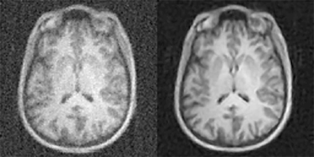
Image: MRI images reconstructed from the same data with conventional approaches (L) and AUTOMAP (R) (Photo courtesy of MGH).
A new technique based on artificial intelligence (AI) and machine learning could enable clinicians to acquire high-quality images from limited data.
Developed at Massachusetts General Hospital (MGH; Boston, USA), the new image manipulation technique, called automated transform by manifold approximation (AUTOMAP), offers a unified framework for image reconstruction by recasting it as a data-driven supervised learning task, which allows mapping between the sensor and the image domain to emerge from an appropriate body of training data. To develop AUTOMAP, the researchers took advantage of the many advances made in neural network models used for AI.
Improvement in graphical processing units (GPUs) that drive the operations also contributed to the powering of image reconstruction algorithms such as AUTOMAP, as they require an immense amount of computation, especially during the training phase. Another factor was the availability of large datasets--known as big data--that are needed to train large neural network models. The overall result is a superior immunity to noise and a reduction in reconstruction artefacts, compared with conventional handcrafted reconstruction methods.
AUTOMAP also offers a number of potential benefits for clinical care, even beyond producing high-quality images in less time with magnetic resonance imaging (MRI) or with lower doses with X-ray, computerized tomography (CT) and positron emission tomography (PET). Because of its processing speed, the technique could help in making real-time decisions about imaging protocols while the patient is still inside the scanner. According to the researchers, the AUTOMAP technique would not have been possible five years ago, or maybe even one year ago. The study was published on March 21, 2018, in Nature.
“The conventional approach to image reconstruction uses a chain of handcrafted signal processing modules that require expert manual parameter tuning’ and often are unable to handle imperfections of the raw data, such as noise,” said lead author Bo Zhu, PhD, of the MGH Martinos Center for Biomedical Imaging. “With AUTOMAP, we've taught imaging systems to 'see' the way humans learn to see after birth, not through directly programming the brain but by promoting neural connections to adapt organically through repeated training on real-world examples.”
“Since AUTOMAP is implemented as a feedforward neural network, the speed of image reconstruction is almost instantaneous, just tens of milliseconds,” said senior author Matt Rosen, PhD, of the center for machine learning at the MGH Martinos. “Our AI approach is showing remarkable improvements in accuracy and noise reduction and thus can advance a wide range of applications. We're incredibly excited to have the opportunity to roll this out into the clinical space where AUTOMAP can work together with inexpensive GPU-accelerated computers to improve clinical imaging and outcomes.”
Deep learning is part of a broader family of AI methods based on learning data representations, as opposed to task specific algorithms. It involves artificial neural network (ANN) algorithms that use a cascade of many layers of nonlinear processing units for feature extraction and transformation, with each successive layer using the output from the previous layer as input to form a hierarchical representation.
Related Links:
Massachusetts General Hospital
Latest General/Advanced Imaging News
- AI-Powered Imaging System Improves Lung Cancer Diagnosis
- AI Model Significantly Enhances Low-Dose CT Capabilities
- Ultra-Low Dose CT Aids Pneumonia Diagnosis in Immunocompromised Patients
- AI Reduces CT Lung Cancer Screening Workload by Almost 80%
- Cutting-Edge Technology Combines Light and Sound for Real-Time Stroke Monitoring
- AI System Detects Subtle Changes in Series of Medical Images Over Time
- New CT Scan Technique to Improve Prognosis and Treatments for Head and Neck Cancers
- World’s First Mobile Whole-Body CT Scanner to Provide Diagnostics at POC
- Comprehensive CT Scans Could Identify Atherosclerosis Among Lung Cancer Patients
- AI Improves Detection of Colorectal Cancer on Routine Abdominopelvic CT Scans
- Super-Resolution Technology Enhances Clinical Bone Imaging to Predict Osteoporotic Fracture Risk
- AI-Powered Abdomen Map Enables Early Cancer Detection
- Deep Learning Model Detects Lung Tumors on CT
- AI Predicts Cardiovascular Risk from CT Scans
- Deep Learning Based Algorithms Improve Tumor Detection in PET/CT Scans
- New Technology Provides Coronary Artery Calcification Scoring on Ungated Chest CT Scans
Channels
Radiography
view channel
World's Largest Class Single Crystal Diamond Radiation Detector Opens New Possibilities for Diagnostic Imaging
Diamonds possess ideal physical properties for radiation detection, such as exceptional thermal and chemical stability along with a quick response time. Made of carbon with an atomic number of six, diamonds... Read more
AI-Powered Imaging Technique Shows Promise in Evaluating Patients for PCI
Percutaneous coronary intervention (PCI), also known as coronary angioplasty, is a minimally invasive procedure where small metal tubes called stents are inserted into partially blocked coronary arteries... Read moreMRI
view channel
AI Tool Tracks Effectiveness of Multiple Sclerosis Treatments Using Brain MRI Scans
Multiple sclerosis (MS) is a condition in which the immune system attacks the brain and spinal cord, leading to impairments in movement, sensation, and cognition. Magnetic Resonance Imaging (MRI) markers... Read more
Ultra-Powerful MRI Scans Enable Life-Changing Surgery in Treatment-Resistant Epileptic Patients
Approximately 360,000 individuals in the UK suffer from focal epilepsy, a condition in which seizures spread from one part of the brain. Around a third of these patients experience persistent seizures... Read more
AI-Powered MRI Technology Improves Parkinson’s Diagnoses
Current research shows that the accuracy of diagnosing Parkinson’s disease typically ranges from 55% to 78% within the first five years of assessment. This is partly due to the similarities shared by Parkinson’s... Read more
Biparametric MRI Combined with AI Enhances Detection of Clinically Significant Prostate Cancer
Artificial intelligence (AI) technologies are transforming the way medical images are analyzed, offering unprecedented capabilities in quantitatively extracting features that go beyond traditional visual... Read moreUltrasound
view channel
AI Identifies Heart Valve Disease from Common Imaging Test
Tricuspid regurgitation is a condition where the heart's tricuspid valve does not close completely during contraction, leading to backward blood flow, which can result in heart failure. A new artificial... Read more
Novel Imaging Method Enables Early Diagnosis and Treatment Monitoring of Type 2 Diabetes
Type 2 diabetes is recognized as an autoimmune inflammatory disease, where chronic inflammation leads to alterations in pancreatic islet microvasculature, a key factor in β-cell dysfunction.... Read moreNuclear Medicine
view channel
Novel PET Imaging Approach Offers Never-Before-Seen View of Neuroinflammation
COX-2, an enzyme that plays a key role in brain inflammation, can be significantly upregulated by inflammatory stimuli and neuroexcitation. Researchers suggest that COX-2 density in the brain could serve... Read more
Novel Radiotracer Identifies Biomarker for Triple-Negative Breast Cancer
Triple-negative breast cancer (TNBC), which represents 15-20% of all breast cancer cases, is one of the most aggressive subtypes, with a five-year survival rate of about 40%. Due to its significant heterogeneity... Read moreImaging IT
view channel
New Google Cloud Medical Imaging Suite Makes Imaging Healthcare Data More Accessible
Medical imaging is a critical tool used to diagnose patients, and there are billions of medical images scanned globally each year. Imaging data accounts for about 90% of all healthcare data1 and, until... Read more
Global AI in Medical Diagnostics Market to Be Driven by Demand for Image Recognition in Radiology
The global artificial intelligence (AI) in medical diagnostics market is expanding with early disease detection being one of its key applications and image recognition becoming a compelling consumer proposition... Read moreIndustry News
view channel
GE HealthCare and NVIDIA Collaboration to Reimagine Diagnostic Imaging
GE HealthCare (Chicago, IL, USA) has entered into a collaboration with NVIDIA (Santa Clara, CA, USA), expanding the existing relationship between the two companies to focus on pioneering innovation in... Read more
Patient-Specific 3D-Printed Phantoms Transform CT Imaging
New research has highlighted how anatomically precise, patient-specific 3D-printed phantoms are proving to be scalable, cost-effective, and efficient tools in the development of new CT scan algorithms... Read more
Siemens and Sectra Collaborate on Enhancing Radiology Workflows
Siemens Healthineers (Forchheim, Germany) and Sectra (Linköping, Sweden) have entered into a collaboration aimed at enhancing radiologists' diagnostic capabilities and, in turn, improving patient care... Read more


 Guided Devices.jpg)


