3D Data Used to Visualize Cardiac Conductive System
|
By MedImaging International staff writers Posted on 14 Aug 2017 |

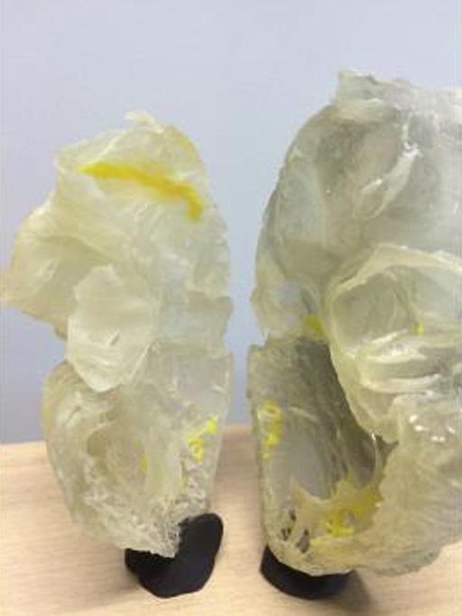
Image: A plastic 3D printed heart highlights the human cardiac conductive system (Photo courtesy of the University of Manchester).
Researchers have discovered new details of how the conductive system of the human heart functions that could help cardiac surgeons repair hearts without damaging healthy tissue.
The results of this pioneering study provide improved and more accurate computer models of the conductive system of the human heart, and the origins of the heartbeat, and could help improve clinicians’ understanding of atrial fibrillation and other common cardiac problems.
The scientists from Liverpool John Moores University (LJMU; Liverpool, UK), The University of Manchester (Manchester, UK), Aarhus University (Aarhus, Denmark), and Newcastle University (Newcastle, UK) published the research findings online in the August 3, 2017, issue of the journal Nature, Scientific Reports.
The scientists soaked post-mortem samples of heart tissue in an iodine solution to enhance visualization of heart tissue in X-Ray images. They then used X-Ray scanners to make 3D images, some of which were so detailed that they showed the boundaries between individual heart cells, and the cellular layout in the tissue.
Professor Jonathan Jarvis, at the LJMU School of Sport and Exercise Sciences, said, "The 3D data makes it much easier to understand the complex relationships between the cardiac conduction system and the rest of the heart. We also use the data to make 3D printed models that are really useful in our discussions with heart doctors, other researchers and patients with heart problems. New strategies to repair or replace the aortic valve must therefore make sure that they do not damage or compress this precious tissue. In future work we will be able to see where the cardiac conduction system runs in hearts that have not formed properly. This will help the surgeons who repair such hearts to design operations that have the least risk of damaging the cardiac conduction system."
Related Links:
Liverpool John Moores University
University of Manchester
Aarhus University
Newcastle University
Latest Imaging IT News
- New Google Cloud Medical Imaging Suite Makes Imaging Healthcare Data More Accessible
- Global AI in Medical Diagnostics Market to Be Driven by Demand for Image Recognition in Radiology
- AI-Based Mammography Triage Software Helps Dramatically Improve Interpretation Process
- Artificial Intelligence (AI) Program Accurately Predicts Lung Cancer Risk from CT Images
- Image Management Platform Streamlines Treatment Plans
- AI-Based Technology for Ultrasound Image Analysis Receives FDA Approval
- AI Technology for Detecting Breast Cancer Receives CE Mark Approval
- Digital Pathology Software Improves Workflow Efficiency
- Patient-Centric Portal Facilitates Direct Imaging Access
- New Workstation Supports Customer-Driven Imaging Workflow
Channels
MRI
view channel
Shorter MRI Exam Effectively Detects Cancer in Dense Breasts
Women with extremely dense breasts face a higher risk of missed breast cancer diagnoses, as dense glandular and fibrous tissue can obscure tumors on mammograms. While breast MRI is recommended for supplemental... Read more
MRI to Replace Painful Spinal Tap for Faster MS Diagnosis
Multiple sclerosis (MS) is a challenging neurological condition to diagnose due to its wide array of symptoms, with not all patients experiencing the same symptoms or at the same intensity, and the disease... Read more
MRI Scans Can Identify Cardiovascular Disease Ten Years in Advance
Cardiovascular disease encompasses various conditions that narrow or block blood vessels, such as heart attacks, strokes, and heart failure. While some individuals are genetically predisposed, lifestyle... Read more
Simple Brain Scan Diagnoses Parkinson's Disease Years Before It Becomes Untreatable
Parkinson's disease (PD) remains a challenging condition to treat, with no known cure. Though therapies have improved over time, and ongoing research focuses on methods to slow or alter the disease’s progression,... Read moreUltrasound
view channel
New Incision-Free Technique Halts Growth of Debilitating Brain Lesions
Cerebral cavernous malformations (CCMs), also known as cavernomas, are abnormal clusters of blood vessels that can grow in the brain, spinal cord, or other parts of the body. While most cases remain asymptomatic,... Read more.jpeg)
AI-Powered Lung Ultrasound Outperforms Human Experts in Tuberculosis Diagnosis
Despite global declines in tuberculosis (TB) rates in previous years, the incidence of TB rose by 4.6% from 2020 to 2023. Early screening and rapid diagnosis are essential elements of the World Health... Read moreNuclear Medicine
view channel
New Imaging Approach Could Reduce Need for Biopsies to Monitor Prostate Cancer
Prostate cancer is the second leading cause of cancer-related death among men in the United States. However, the majority of older men diagnosed with prostate cancer have slow-growing, low-risk forms of... Read more
Novel Radiolabeled Antibody Improves Diagnosis and Treatment of Solid Tumors
Interleukin-13 receptor α-2 (IL13Rα2) is a cell surface receptor commonly found in solid tumors such as glioblastoma, melanoma, and breast cancer. It is minimally expressed in normal tissues, making it... Read moreGeneral/Advanced Imaging
view channel
First-Of-Its-Kind Wearable Device Offers Revolutionary Alternative to CT Scans
Currently, patients with conditions such as heart failure, pneumonia, or respiratory distress often require multiple imaging procedures that are intermittent, disruptive, and involve high levels of radiation.... Read more
AI-Based CT Scan Analysis Predicts Early-Stage Kidney Damage Due to Cancer Treatments
Radioligand therapy, a form of targeted nuclear medicine, has recently gained attention for its potential in treating specific types of tumors. However, one of the potential side effects of this therapy... Read moreImaging IT
view channel
New Google Cloud Medical Imaging Suite Makes Imaging Healthcare Data More Accessible
Medical imaging is a critical tool used to diagnose patients, and there are billions of medical images scanned globally each year. Imaging data accounts for about 90% of all healthcare data1 and, until... Read more
Global AI in Medical Diagnostics Market to Be Driven by Demand for Image Recognition in Radiology
The global artificial intelligence (AI) in medical diagnostics market is expanding with early disease detection being one of its key applications and image recognition becoming a compelling consumer proposition... Read moreIndustry News
view channel
GE HealthCare and NVIDIA Collaboration to Reimagine Diagnostic Imaging
GE HealthCare (Chicago, IL, USA) has entered into a collaboration with NVIDIA (Santa Clara, CA, USA), expanding the existing relationship between the two companies to focus on pioneering innovation in... Read more
Patient-Specific 3D-Printed Phantoms Transform CT Imaging
New research has highlighted how anatomically precise, patient-specific 3D-printed phantoms are proving to be scalable, cost-effective, and efficient tools in the development of new CT scan algorithms... Read more
Siemens and Sectra Collaborate on Enhancing Radiology Workflows
Siemens Healthineers (Forchheim, Germany) and Sectra (Linköping, Sweden) have entered into a collaboration aimed at enhancing radiologists' diagnostic capabilities and, in turn, improving patient care... Read more


