Study Suggests Teenage Obesity May Lead to Permanent Bone Loss
|
By Andrew Deutsch Posted on 22 Nov 2016 |

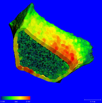
Image: A thickness map of the radius bone of the forearm, which was acquired using a SCANCO Medical Xtreme CT scanner (Photo courtesy of RSNA).
Researchers have shown that obesity in adolescents affects bone density and could increase the risk of bone fractures in later life.
Obesity is a major problem in many countries and is commonly associated with cardiovascular disease, and diabetes. The goal of the researchers is to try and find how obesity in adolescents affects bone structure.
Twenty-three adolescents with a mean Body Mass Index (BMI) of 44 kg/m2, and with a mean age of 17 years took part in the study. The researchers used 3D High Resolution Peripheral Quantitative Computed Tomography (HR-pQCT) exams to measure bone microarchitecture, and mineral density in the arms and legs of the study participants. The scans enabled the researchers to study the structure of a bone in the forearm called the distal radius. In addition, the study participants underwent dual-energy X-Ray Absorptiometry (DXA) exams to quantify lean mass, and visceral fat mass. The research was presented at the annual Radiological Society of North America (RSNA2016) meeting.
The study results showed a positive association between BMI and cortical bone thickness and area, and cortical bone porosity. There was also a positive association between lean mass and trabecular density, bone volume, and integrity. The researchers concluded that a high amount of visceral fat combined with a low amount of muscle mass, was a risk factor for weakened bone structure in adolescents.
Lead author of the study, radiologist Miriam A. Bredella, MD, Massachusetts General Hospital (Boston. MA, USA), said, "While obesity was previously believed to be protective of bone health, recent studies have shown a higher incidence of forearm fractures in obese youths. Adolescence is the time where we accrue our peak bone mass, so bone loss during this time is a serious problem. We know from other chronic states that lead to bone loss in adolescence, such as anorexia nervosa, that increased fracture risk persists in adulthood, even after normalization of body weight. Therefore, it is important to address this problem early on. In addition, vitamin D, which is important for bone health, is soluble in adipose tissue and gets trapped within fat cells. The best way to prevent bone loss is a healthy diet that contains adequate amounts of calcium and vitamin D, along with sufficient exercise, as we have shown in our study that muscle mass is good for bone health."
Related Links:
Massachusetts General Hospital
Latest Radiography News
- AI-Powered Imaging Technique Shows Promise in Evaluating Patients for PCI
- Higher Chest X-Ray Usage Catches Lung Cancer Earlier and Improves Survival
- AI-Powered Mammograms Predict Cardiovascular Risk
- Generative AI Model Significantly Reduces Chest X-Ray Reading Time
- AI-Powered Mammography Screening Boosts Cancer Detection in Single-Reader Settings
- Photon Counting Detectors Promise Fast Color X-Ray Images
- AI Can Flag Mammograms for Supplemental MRI
- 3D CT Imaging from Single X-Ray Projection Reduces Radiation Exposure
- AI Method Accurately Predicts Breast Cancer Risk by Analyzing Multiple Mammograms
- Printable Organic X-Ray Sensors Could Transform Treatment for Cancer Patients
- Highly Sensitive, Foldable Detector to Make X-Rays Safer
- Novel Breast Cancer Screening Technology Could Offer Superior Alternative to Mammogram
- Artificial Intelligence Accurately Predicts Breast Cancer Years Before Diagnosis
- AI-Powered Chest X-Ray Detects Pulmonary Nodules Three Years Before Lung Cancer Symptoms
- AI Model Identifies Vertebral Compression Fractures in Chest Radiographs
- Advanced 3D Mammography Detects More Breast Cancers
Channels
MRI
view channel
Ultra-Powerful MRI Scans Enable Life-Changing Surgery in Treatment-Resistant Epileptic Patients
Approximately 360,000 individuals in the UK suffer from focal epilepsy, a condition in which seizures spread from one part of the brain. Around a third of these patients experience persistent seizures... Read more
AI-Powered MRI Technology Improves Parkinson’s Diagnoses
Current research shows that the accuracy of diagnosing Parkinson’s disease typically ranges from 55% to 78% within the first five years of assessment. This is partly due to the similarities shared by Parkinson’s... Read more
Biparametric MRI Combined with AI Enhances Detection of Clinically Significant Prostate Cancer
Artificial intelligence (AI) technologies are transforming the way medical images are analyzed, offering unprecedented capabilities in quantitatively extracting features that go beyond traditional visual... Read more
First-Of-Its-Kind AI-Driven Brain Imaging Platform to Better Guide Stroke Treatment Options
Each year, approximately 800,000 people in the U.S. experience strokes, with marginalized and minoritized groups being disproportionately affected. Strokes vary in terms of size and location within the... Read moreUltrasound
view channel
Smart Ultrasound-Activated Immune Cells Destroy Cancer Cells for Extended Periods
Chimeric antigen receptor (CAR) T-cell therapy has emerged as a highly promising cancer treatment, especially for bloodborne cancers like leukemia. This highly personalized therapy involves extracting... Read more
Tiny Magnetic Robot Takes 3D Scans from Deep Within Body
Colorectal cancer ranks as one of the leading causes of cancer-related mortality worldwide. However, when detected early, it is highly treatable. Now, a new minimally invasive technique could significantly... Read more
High Resolution Ultrasound Speeds Up Prostate Cancer Diagnosis
Each year, approximately one million prostate cancer biopsies are conducted across Europe, with similar numbers in the USA and around 100,000 in Canada. Most of these biopsies are performed using MRI images... Read more
World's First Wireless, Handheld, Whole-Body Ultrasound with Single PZT Transducer Makes Imaging More Accessible
Ultrasound devices play a vital role in the medical field, routinely used to examine the body's internal tissues and structures. While advancements have steadily improved ultrasound image quality and processing... Read moreNuclear Medicine
view channel
Novel PET Imaging Approach Offers Never-Before-Seen View of Neuroinflammation
COX-2, an enzyme that plays a key role in brain inflammation, can be significantly upregulated by inflammatory stimuli and neuroexcitation. Researchers suggest that COX-2 density in the brain could serve... Read more
Novel Radiotracer Identifies Biomarker for Triple-Negative Breast Cancer
Triple-negative breast cancer (TNBC), which represents 15-20% of all breast cancer cases, is one of the most aggressive subtypes, with a five-year survival rate of about 40%. Due to its significant heterogeneity... Read moreGeneral/Advanced Imaging
view channel
AI-Powered Imaging System Improves Lung Cancer Diagnosis
Given the need to detect lung cancer at earlier stages, there is an increasing need for a definitive diagnostic pathway for patients with suspicious pulmonary nodules. However, obtaining tissue samples... Read more
AI Model Significantly Enhances Low-Dose CT Capabilities
Lung cancer remains one of the most challenging diseases, making early diagnosis vital for effective treatment. Fortunately, advancements in artificial intelligence (AI) are revolutionizing lung cancer... Read moreImaging IT
view channel
New Google Cloud Medical Imaging Suite Makes Imaging Healthcare Data More Accessible
Medical imaging is a critical tool used to diagnose patients, and there are billions of medical images scanned globally each year. Imaging data accounts for about 90% of all healthcare data1 and, until... Read more
Global AI in Medical Diagnostics Market to Be Driven by Demand for Image Recognition in Radiology
The global artificial intelligence (AI) in medical diagnostics market is expanding with early disease detection being one of its key applications and image recognition becoming a compelling consumer proposition... Read moreIndustry News
view channel
GE HealthCare and NVIDIA Collaboration to Reimagine Diagnostic Imaging
GE HealthCare (Chicago, IL, USA) has entered into a collaboration with NVIDIA (Santa Clara, CA, USA), expanding the existing relationship between the two companies to focus on pioneering innovation in... Read more
Patient-Specific 3D-Printed Phantoms Transform CT Imaging
New research has highlighted how anatomically precise, patient-specific 3D-printed phantoms are proving to be scalable, cost-effective, and efficient tools in the development of new CT scan algorithms... Read more
Siemens and Sectra Collaborate on Enhancing Radiology Workflows
Siemens Healthineers (Forchheim, Germany) and Sectra (Linköping, Sweden) have entered into a collaboration aimed at enhancing radiologists' diagnostic capabilities and, in turn, improving patient care... Read more


