MRI Measures Knee Geometry and Its Role in Severe Knee Injuries
|
By MedImaging International staff writers Posted on 06 Oct 2014 |

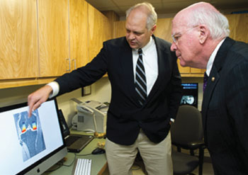
Image: University of Vermont professor of musculoskeletal research Bruce Beynnon, PhD, left, describes how the University of Vermont uses their research MRI machine in knee injury research to US Senator Patrick Leahy (Photo courtesy of COM Design & Photography).
Researchers are examining multiple factors such as the size of the knee’s femoral notch to attempt and clarify why some people are at greater risk for injury than others.
The moving of an athlete’s body relies on a coordinated response of joints, ligaments, bones, and tendons, putting the geometry of a key part like a knee joint to the test. However, it is more than just a footfall mistake at the root cause of one of the most devastating of sports injuries: the anterior cruciate ligament (ACL) tear. In reality, size—of the femoral notch that is situated at the center of the knee joint—and volume of the ACL combine to influence the risk of suffering a noncontact ACL injury. Additional geometric characteristics of the knee, such as the slope of the articular surfaces, are involved with risk of injury.
Several recent studies—including a highly-controlled study of first-time ACL injuries in Vermont high school and college athletic team members—conducted by University of Vermont (UVM; Burlington, USA) professor of musculoskeletal research Bruce Beynnon, PhD, and colleagues. With only 200,000 to 300,000 injuries yearly, ACL injuries are far less common than ankle ligament injuries, which number more than two million injuries in the United States alone yearly. However, ACL injuries can end sports careers and are proven to lead to the early onset of osteoarthritis.
“It’s a concern because its highly likely that an individual that suffers this injury will progress to end-stage arthritis in 15 years and the only solution at that point in time—knee joint replacement—only lasts about 15 to 20 years in an active individual,” explained Prof. Beynnon.
While the rate of ACL injuries across the United States have not changed over time, according to Prof. Beynnon, he and his team have looked at and continue to research the many variables at play when this injury takes place. In the study, published August 2014 in the American Journal of Sports Medicine, he and colleagues have “very accurately characterized the incidence rate and magnitude of this problem in Vermont,” stated Prof. Beynnon.
The researchers examined 88 student athletes—27 male and 61 female—who suffered first-time, noncontact ACL injuries during the study and compared their measurements—collected using magnetic resonance imaging (MRI) images of their knees—to a non-injured control group of 88 athletes (same male-female breakdown) from the same teams, with the same extrinsic factors, like environment, playing surface, training, footwear, level of competition, and coaching.
These measurements led them to look at the point where the ACL is positioned in the center of the knee—the femoral notch—where they measured its width, as well as ACL volume utilizing MRI technology in the University of Vermont’s MRI Center for Biomedical Imaging. One of the findings they discovered is that the risk of injury increased as the size of the femoral notch and ACL decreases.
“Prognostic studies are designed to identify who is likely to suffer injury so we can target injury prevention programs at them, and determine why they are at increased risk so we can inform the development of the programs to reduce the risk of injury and re-injuries of the same kind,” noted Dr. Beynnon.
In a parallel five-year epidemiologic study, also published in the August 2014 American Journal of Sports Medicine, the researchers reported on “The Effects of Level of Competition, Sport, and Sex on the Incidence of First-Time Noncontact Anterior Cruciate Ligament Injury.”
From the data gathered from a total of 38 institutions located throughout the US state of Vermont, colleges reported 48 ACL injuries during the sport seasons studied, while high schools reported 53 injuries. The researchers learned that college athletes had a significantly higher ACL injury risk than high school athletes—approximately two-fold—and that female athletes were two times more at risk for ACL injuries than males. In comparison to athletes taking part in Lacrosse, risk of ACL injury was substantially greater among those participating in soccer and rugby.
In their conclusion, the study’s authors stated, “An athlete’s risk of having a first-time noncontact ACL injury is independently influenced by level of competition, the participant’s sex, and type of sport they participate in, and there are no interactions between their effects. Female college athletes have the highest risk of having a first-time noncontact ACL injury among the groups studied. The first step is to establish the athletes most at risk; targeting interventions comes second.”
The investigators are currently conducting a multivariable study that has 109 participants. They are examining the role of a range of diverse factors to try to additionally identify those at increased risk for ACL injury. “It’s not just biomechanics, it’s biomechanics and biology,” Prof. Beynnon stated, adding that family history—a genetic link—could be the driver of such variables as problems with ACL collagen synthesis and degradation, joint laxity, exercise and diet, body mass index, and an individual’s inflammatory response.
Prof. Beynnon hopes, in five to 10 years, his group can repeat their epidemiologic study to validate their findings, evaluate the high school and college athlete model on a different, independent sample of individuals, and is considering developing an intervention study. “Primary prevention is the ultimate goal,” he said. “We want to reduce the risk of injury and burden of disease for this young age group.”
Related Links:
University of Vermont
Latest MRI News
- MRI to Replace Painful Spinal Tap for Faster MS Diagnosis
- MRI Scans Can Identify Cardiovascular Disease Ten Years in Advance
- Simple Brain Scan Diagnoses Parkinson's Disease Years Before It Becomes Untreatable
- Cutting-Edge MRI Technology to Revolutionize Diagnosis of Common Heart Problem
- New MRI Technique Reveals True Heart Age to Prevent Attacks and Strokes
- AI Tool Predicts Relapse of Pediatric Brain Cancer from Brain MRI Scans
- AI Tool Tracks Effectiveness of Multiple Sclerosis Treatments Using Brain MRI Scans
- Ultra-Powerful MRI Scans Enable Life-Changing Surgery in Treatment-Resistant Epileptic Patients
- AI-Powered MRI Technology Improves Parkinson’s Diagnoses
- Biparametric MRI Combined with AI Enhances Detection of Clinically Significant Prostate Cancer
- First-Of-Its-Kind AI-Driven Brain Imaging Platform to Better Guide Stroke Treatment Options
- New Model Improves Comparison of MRIs Taken at Different Institutions
- Groundbreaking New Scanner Sees 'Previously Undetectable' Cancer Spread
- First-Of-Its-Kind Tool Analyzes MRI Scans to Measure Brain Aging
- AI-Enhanced MRI Images Make Cancerous Breast Tissue Glow
- AI Model Automatically Segments MRI Images
Channels
Radiography
view channel
Machine Learning Algorithm Identifies Cardiovascular Risk from Routine Bone Density Scans
A new study published in the Journal of Bone and Mineral Research reveals that an automated machine learning program can predict the risk of cardiovascular events and falls or fractures by analyzing bone... Read more
AI Improves Early Detection of Interval Breast Cancers
Interval breast cancers, which occur between routine screenings, are easier to treat when detected earlier. Early detection can reduce the need for aggressive treatments and improve the chances of better outcomes.... Read more
World's Largest Class Single Crystal Diamond Radiation Detector Opens New Possibilities for Diagnostic Imaging
Diamonds possess ideal physical properties for radiation detection, such as exceptional thermal and chemical stability along with a quick response time. Made of carbon with an atomic number of six, diamonds... Read moreUltrasound
view channel
New Incision-Free Technique Halts Growth of Debilitating Brain Lesions
Cerebral cavernous malformations (CCMs), also known as cavernomas, are abnormal clusters of blood vessels that can grow in the brain, spinal cord, or other parts of the body. While most cases remain asymptomatic,... Read more.jpeg)
AI-Powered Lung Ultrasound Outperforms Human Experts in Tuberculosis Diagnosis
Despite global declines in tuberculosis (TB) rates in previous years, the incidence of TB rose by 4.6% from 2020 to 2023. Early screening and rapid diagnosis are essential elements of the World Health... Read moreNuclear Medicine
view channel
New Imaging Approach Could Reduce Need for Biopsies to Monitor Prostate Cancer
Prostate cancer is the second leading cause of cancer-related death among men in the United States. However, the majority of older men diagnosed with prostate cancer have slow-growing, low-risk forms of... Read more
Novel Radiolabeled Antibody Improves Diagnosis and Treatment of Solid Tumors
Interleukin-13 receptor α-2 (IL13Rα2) is a cell surface receptor commonly found in solid tumors such as glioblastoma, melanoma, and breast cancer. It is minimally expressed in normal tissues, making it... Read moreGeneral/Advanced Imaging
view channel
First-Of-Its-Kind Wearable Device Offers Revolutionary Alternative to CT Scans
Currently, patients with conditions such as heart failure, pneumonia, or respiratory distress often require multiple imaging procedures that are intermittent, disruptive, and involve high levels of radiation.... Read more
AI-Based CT Scan Analysis Predicts Early-Stage Kidney Damage Due to Cancer Treatments
Radioligand therapy, a form of targeted nuclear medicine, has recently gained attention for its potential in treating specific types of tumors. However, one of the potential side effects of this therapy... Read moreImaging IT
view channel
New Google Cloud Medical Imaging Suite Makes Imaging Healthcare Data More Accessible
Medical imaging is a critical tool used to diagnose patients, and there are billions of medical images scanned globally each year. Imaging data accounts for about 90% of all healthcare data1 and, until... Read more
Global AI in Medical Diagnostics Market to Be Driven by Demand for Image Recognition in Radiology
The global artificial intelligence (AI) in medical diagnostics market is expanding with early disease detection being one of its key applications and image recognition becoming a compelling consumer proposition... Read moreIndustry News
view channel
GE HealthCare and NVIDIA Collaboration to Reimagine Diagnostic Imaging
GE HealthCare (Chicago, IL, USA) has entered into a collaboration with NVIDIA (Santa Clara, CA, USA), expanding the existing relationship between the two companies to focus on pioneering innovation in... Read more
Patient-Specific 3D-Printed Phantoms Transform CT Imaging
New research has highlighted how anatomically precise, patient-specific 3D-printed phantoms are proving to be scalable, cost-effective, and efficient tools in the development of new CT scan algorithms... Read more
Siemens and Sectra Collaborate on Enhancing Radiology Workflows
Siemens Healthineers (Forchheim, Germany) and Sectra (Linköping, Sweden) have entered into a collaboration aimed at enhancing radiologists' diagnostic capabilities and, in turn, improving patient care... Read more


