New Cardiology Imaging Tools Include MRI Myocardial Tissue Quantification to Help Fight Cardiovascular Disease
|
By MedImaging International staff writers Posted on 10 Sep 2014 |

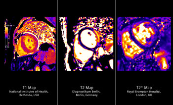
Image: T1, T2 and T2 myocardial tissue quantification in one solution, on the fly. Based on HeartFreeze Inline Motion Correction (Siemens unique), MyoMaps1 provides pixel-based myocardial quantification. Global, diffuse, myocardial pathologies can be better detected (T1 Map), or better depict cardiac edema (T2 Map) and improve early detection of iron overload (T2 Map) (Photo courtesy of Siemens Healthcare).
New imaging tools have been designed for a more precise diagnosis of cardiovascular diseases using computed tomography (CT), magnetic resonance imaging (MRI), and molecular imaging, as well as utilizing a universal angiography system with sophisticated features for cardiology.
An MRI application has been designed for myocardial tissue quantification, in addition to precise and effective myocardial perfusion due to a new application for CT systems. Siemens Healthcare (Erlangen, Germany) presented its latest cardiology imaging technology at the Congress of the European Society for Cardiology (ESC), held August 30 to September 3, 2014, in Barcelona (Spain).
Early detection of heart failure with MRI cardiac magnetic resonance imaging (CMR) provides detailed information about the morphology and function of the heart. Current ESC guidelines consider CMR to be the gold standard for the diagnosis and treatment of acute and chronic heart failure. This is largely due to MRI’s accuracy in measuring heart volume, mass, and wall motion, including evaluation of ischemia and viability.
The MyoMaps application not only enables visual diagnosis but also offers physical quantification of myocardial tissue with MyoMaps. The tool makes it possible to record microscopic changes in myocardial tissue and represent them as pixel-based, colored images. This enables the diagnosis and quantification of diffuse myocardial pathologies, scar tissue, and edemas in the very early stages of the disease. It also allows for early detection and quantification of iron overload, which can result in heart failure.
With these findings, physicians can diagnose and begin treating heart patients earlier than before and improve the course of disease. MyoMaps is now available for the 1.5-Tesla scanner Magnetom Aera and the 3.0-Tesla scanner Magnetom Skyra, and will be available for more scanners in the future.
Using CT to evaluate the total range of myocardial perfusion, one of the developments in computed tomography highlighted by Siemens at this year’s ESC congress is the Syngo.CT cardiac function-enhancement application for myocardial perfusion. With this application, both CT angiography and functional assessment of coronary lesions, using perfusion imaging, can be performed using only one modality. This means more convenience for the patients, offers a faster approach to diagnosis for the physician, and saves time. Combined with the Somatom Force CT scanner, it is very easy to incorporate the tool into daily routine, since it offers substantially improved spatial coverage of the heart.
CT angiography is used to examine the coronary arteries and identify stenosis, indicating risks to myocardial blood supply. The evaluation of myocardial perfusion provides physicians with important additional data to enable them to decide whether the patient is suffering from a hemodynamically relevant stenosis requiring intervention in the cath lab. Precise and insightful diagnostics are particularly important in the case of patients with moderately severe stenosis, where a diagnosis based only on CT angiography findings is not possible.
Physicians can achieve this evaluation using Syngo.CT Cardiac function-Enhancement and an examination using the Somatom Force or Somatom Definition Flash. They can choose from the entire range of myocardial perfusion, regardless of whether the process involves first-pass enhancement for the qualitative identification of perfusion defects, late enhancement to diagnose tissue affected by scar lesions, or the quantitative evaluation of myocardial blood flow via dynamic myocardial perfusion.
Three new applications for the Biograph mCT Flow positron emission tomography-computed tomography (PET-CT) scanner from Siemens Healthcare will also be featured at this year’s ESC congress. “Phase matching can be used with PET/CT examinations to enhance quantification and image quality. Identical displays, for instance, are automatically provided for the cardiac phase in both PET and CT measurement, enabling physicians to compare both datasets directly against each other.
The Smart auto cardiac registration application automatically aligns the PET and CT images based on anatomical landmarks, providing for the correct values for PET attenuation correction at all times, even if the CT and PET data are acquired in different time points. This previously had to be done manually, which meant more time was needed and the results were less reproducible.
HD•Cardiac is a new application that enables respiratory motion management on PET cardiac scans without the need for an external respiratory trigger device. This device had earlier been needed to synchronize the acquisition with the respiratory cycle. Now, however, electrocardiography (ECG) gating and respiratory gating are performed together. There is no need for an additional respiratory gating unit next to the ECG, because the movement of the heart caused by breathing can be corrected by the PET data itself. The result is reduced respiratory motion blur, improved visualization of PET images and less complicated patient positioning.
Minimally invasive procedures are becoming more and more popular world-wide. In cardiology, these mainly include dilation of narrowed coronary arteries (coronary stenosis) or revascularization of peripheral artery disease, and implanting pacemakers. Siemens Healthcare developed Artis one angiography system with advanced features for all of these routine interventions, and will showcase it at the ESC 2014 congress.
The angiography system is similar in positioning flexibility to a ceiling-mounted system, while requiring much less space: Artis one needs only 25 square meters, compared to the usual 45 square meters for ceiling-mounted systems. It has multiple axes that can be moved independently of each other, enabling physicians and clinical staff to select the most appropriate system position with ease to suit every procedure, regardless whether the physician stands on the patient’s right, as in the case of a coronary examination, or on the left, for pacemaker implantations.
The inclusion of Clearstent Live provides Artis one with a feature that was previously available only for the premium product family Artis Q and Artis Q.zen. This cardiology application enables the physician to mask out movement of the beating heart in order to place the stent in precisely the right position. The new HeartSweep functionality makes use of the system’s double axis rotation and enables to capture all cardiac standard projections in one movement of the C-arm. This can be done in only five seconds and with only one injection of contrast agent.
Related Links:
Siemens Healthcare
Latest MRI News
- Ultra-Powerful MRI Scans Enable Life-Changing Surgery in Treatment-Resistant Epileptic Patients
- AI-Powered MRI Technology Improves Parkinson’s Diagnoses
- Biparametric MRI Combined with AI Enhances Detection of Clinically Significant Prostate Cancer
- First-Of-Its-Kind AI-Driven Brain Imaging Platform to Better Guide Stroke Treatment Options
- New Model Improves Comparison of MRIs Taken at Different Institutions
- Groundbreaking New Scanner Sees 'Previously Undetectable' Cancer Spread
- First-Of-Its-Kind Tool Analyzes MRI Scans to Measure Brain Aging
- AI-Enhanced MRI Images Make Cancerous Breast Tissue Glow
- AI Model Automatically Segments MRI Images
- New Research Supports Routine Brain MRI Screening in Asymptomatic Late-Stage Breast Cancer Patients
- Revolutionary Portable Device Performs Rapid MRI-Based Stroke Imaging at Patient's Bedside
- AI Predicts After-Effects of Brain Tumor Surgery from MRI Scans
- MRI-First Strategy for Prostate Cancer Detection Proven Safe
- First-Of-Its-Kind 10' x 48' Mobile MRI Scanner Transforms User and Patient Experience
- New Model Makes MRI More Accurate and Reliable
- New Scan Method Shows Effects of Treatment on Lung Function in Real Time
Channels
Radiography
view channel
AI-Powered Imaging Technique Shows Promise in Evaluating Patients for PCI
Percutaneous coronary intervention (PCI), also known as coronary angioplasty, is a minimally invasive procedure where small metal tubes called stents are inserted into partially blocked coronary arteries... Read more
Higher Chest X-Ray Usage Catches Lung Cancer Earlier and Improves Survival
Lung cancer continues to be the leading cause of cancer-related deaths worldwide. While advanced technologies like CT scanners play a crucial role in detecting lung cancer, more accessible and affordable... Read moreUltrasound
view channel
Smart Ultrasound-Activated Immune Cells Destroy Cancer Cells for Extended Periods
Chimeric antigen receptor (CAR) T-cell therapy has emerged as a highly promising cancer treatment, especially for bloodborne cancers like leukemia. This highly personalized therapy involves extracting... Read more
Tiny Magnetic Robot Takes 3D Scans from Deep Within Body
Colorectal cancer ranks as one of the leading causes of cancer-related mortality worldwide. However, when detected early, it is highly treatable. Now, a new minimally invasive technique could significantly... Read more
High Resolution Ultrasound Speeds Up Prostate Cancer Diagnosis
Each year, approximately one million prostate cancer biopsies are conducted across Europe, with similar numbers in the USA and around 100,000 in Canada. Most of these biopsies are performed using MRI images... Read more
World's First Wireless, Handheld, Whole-Body Ultrasound with Single PZT Transducer Makes Imaging More Accessible
Ultrasound devices play a vital role in the medical field, routinely used to examine the body's internal tissues and structures. While advancements have steadily improved ultrasound image quality and processing... Read moreNuclear Medicine
view channel
Novel PET Imaging Approach Offers Never-Before-Seen View of Neuroinflammation
COX-2, an enzyme that plays a key role in brain inflammation, can be significantly upregulated by inflammatory stimuli and neuroexcitation. Researchers suggest that COX-2 density in the brain could serve... Read more
Novel Radiotracer Identifies Biomarker for Triple-Negative Breast Cancer
Triple-negative breast cancer (TNBC), which represents 15-20% of all breast cancer cases, is one of the most aggressive subtypes, with a five-year survival rate of about 40%. Due to its significant heterogeneity... Read moreGeneral/Advanced Imaging
view channel
AI-Powered Imaging System Improves Lung Cancer Diagnosis
Given the need to detect lung cancer at earlier stages, there is an increasing need for a definitive diagnostic pathway for patients with suspicious pulmonary nodules. However, obtaining tissue samples... Read more
AI Model Significantly Enhances Low-Dose CT Capabilities
Lung cancer remains one of the most challenging diseases, making early diagnosis vital for effective treatment. Fortunately, advancements in artificial intelligence (AI) are revolutionizing lung cancer... Read moreImaging IT
view channel
New Google Cloud Medical Imaging Suite Makes Imaging Healthcare Data More Accessible
Medical imaging is a critical tool used to diagnose patients, and there are billions of medical images scanned globally each year. Imaging data accounts for about 90% of all healthcare data1 and, until... Read more
Global AI in Medical Diagnostics Market to Be Driven by Demand for Image Recognition in Radiology
The global artificial intelligence (AI) in medical diagnostics market is expanding with early disease detection being one of its key applications and image recognition becoming a compelling consumer proposition... Read moreIndustry News
view channel
GE HealthCare and NVIDIA Collaboration to Reimagine Diagnostic Imaging
GE HealthCare (Chicago, IL, USA) has entered into a collaboration with NVIDIA (Santa Clara, CA, USA), expanding the existing relationship between the two companies to focus on pioneering innovation in... Read more
Patient-Specific 3D-Printed Phantoms Transform CT Imaging
New research has highlighted how anatomically precise, patient-specific 3D-printed phantoms are proving to be scalable, cost-effective, and efficient tools in the development of new CT scan algorithms... Read more
Siemens and Sectra Collaborate on Enhancing Radiology Workflows
Siemens Healthineers (Forchheim, Germany) and Sectra (Linköping, Sweden) have entered into a collaboration aimed at enhancing radiologists' diagnostic capabilities and, in turn, improving patient care... Read more


