Advances in Imaging Software Reduce Patient Wait Times and Helps Hospital Staff Increase Efficiency
|
By MedImaging International staff writers Posted on 27 Jan 2014 |

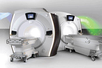
Image: The DV24.0 Continuum Pak allows productivity enhancements of up to 30% compared to earlier versions, increasing efficiency by reducing the number of mouse clicks for technologists (Photo courtesy of GE Healthcare).
Magnetic resonance imaging (MRI), nuclear medicine, radiography and fluoroscopy software has been developed to improve clinical capabilities, customization, lower costs, and streamline operational results.
GE Healthcare, a unit of General Electric Company (Chalfont St. Giles, UK), has developed four software systems to provide improved clinical capabilities, productivity, and diagnostic confidence for clinicians. Up-to-date and effective software is critical to quality care for the patient and practicality for the clinician. GE Healthcare recently announced a USD 2 billion investment in software development that is focused on enhancing asset performance, improving hospital operations management, improving clinical effectiveness and optimizing care across entire populations. “The personalized workflow and unique new clinical applications will give clinicians the information they demand with speed, convenience, and flexibility,” said Nathan Hermony, general manager for GE Healthcare’s nuclear medicine business.
An innovative MRI software platform features novel applications such as the Silent Scan. This neuro acquisition tool makes the sound of an MR scan as quiet as a whisper. “The DV24.0 Continuum Pak upgrade that includes Silent Scan is, in my opinion, the most innovative approach in MRI in the last few years that will make a significant contribution in the chain of diagnosis,” said Jean-Marc Pinon, manager of MR and computed tomography (CT) at CHR Laennec (Creil, France).
DV24.0 Continuum Pak is available on the Optima MR450w, Optima MR450w with GEM, Discovery MR750, and Discovery MR750w with GEM scanners. With DV24.0, productivity enhancements of up to 30% compared to earlier versions are now possible with new features such as the eXpress PreScan and Workflow 2.0, increasing efficiency by reducing the number of mouse clicks for technologists. DV24.0 also features enhancements to increase diagnostic effectiveness. By evaluating the top clinical needs and trends, the GE MRI system has improved 3D imaging through a real-time motion correction technique called PROMO that automatically compensates for head motion.
Moreover, DV24.0, with FOCUS, has the ability to optimize for image quality and signal to noise. FOCUS provides high-resolution, organ-specific diffusion-weighted imaging (DWI) and diffusion tensor imaging (DTI) for a small field of view. Mavric SL, a new technique designed for imaging the joints of patients with MR-conditional implants, is also featured in the DV24.0.
GE Healthcare has expanded its suite of enterprise imaging systems to help lower information technology (IT) costs, improve clinician productivity, expand networks of care, and enhance diagnostic effectiveness. The Centricity 360 systems connect unaffiliated clinicians and patients through a professional online collaboration tool and gives them secure on demand access to imaging applications. Centricity 360 is the newest addition to GE’s Predictivity solutions, which uses the Industrial Internet to help industrial organizations achieve zero unplanned downtime and optimal productivity. Additionally, GE Healthcare’s enterprise imaging systems include Centricity picture archiving and communication system (PACS) with Universal Viewer and Centricity Clinical Archive.
GE Healthcare’s iCenter provides instant access to key data such as asset status, location, maintenance history, and use and planning, which helps enable data-driven decisions and improved operational results. Information is now easier to sift through with a more dynamic, customizable and colorful user interface. Features include fewer clicks, a built-in analytics engine for more visual and intuitive data depiction, user security enhancements, and integrated guided tours. iCenter is the platform for future GE Healthcare’s online service applications.
Related Links:
GE Healthcare
Latest Nuclear Medicine News
- Novel Radiolabeled Antibody Improves Diagnosis and Treatment of Solid Tumors
- Novel PET Imaging Approach Offers Never-Before-Seen View of Neuroinflammation
- Novel Radiotracer Identifies Biomarker for Triple-Negative Breast Cancer
- Innovative PET Imaging Technique to Help Diagnose Neurodegeneration
- New Molecular Imaging Test to Improve Lung Cancer Diagnosis
- Novel PET Technique Visualizes Spinal Cord Injuries to Predict Recovery
- Next-Gen Tau Radiotracers Outperform FDA-Approved Imaging Agents in Detecting Alzheimer’s
- Breakthrough Method Detects Inflammation in Body Using PET Imaging
- Advanced Imaging Reveals Hidden Metastases in High-Risk Prostate Cancer Patients
- Combining Advanced Imaging Technologies Offers Breakthrough in Glioblastoma Treatment
- New Molecular Imaging Agent Accurately Identifies Crucial Cancer Biomarker
- New Scans Light Up Aggressive Tumors for Better Treatment
- AI Stroke Brain Scan Readings Twice as Accurate as Current Method
- AI Analysis of PET/CT Images Predicts Side Effects of Immunotherapy in Lung Cancer
- New Imaging Agent to Drive Step-Change for Brain Cancer Imaging
- Portable PET Scanner to Detect Earliest Stages of Alzheimer’s Disease
Channels
Radiography
view channel
AI Improves Early Detection of Interval Breast Cancers
Interval breast cancers, which occur between routine screenings, are easier to treat when detected earlier. Early detection can reduce the need for aggressive treatments and improve the chances of better outcomes.... Read more
World's Largest Class Single Crystal Diamond Radiation Detector Opens New Possibilities for Diagnostic Imaging
Diamonds possess ideal physical properties for radiation detection, such as exceptional thermal and chemical stability along with a quick response time. Made of carbon with an atomic number of six, diamonds... Read moreMRI
view channel
Cutting-Edge MRI Technology to Revolutionize Diagnosis of Common Heart Problem
Aortic stenosis is a common and potentially life-threatening heart condition. It occurs when the aortic valve, which regulates blood flow from the heart to the rest of the body, becomes stiff and narrow.... Read more
New MRI Technique Reveals True Heart Age to Prevent Attacks and Strokes
Heart disease remains one of the leading causes of death worldwide. Individuals with conditions such as diabetes or obesity often experience accelerated aging of their hearts, sometimes by decades.... Read more
AI Tool Predicts Relapse of Pediatric Brain Cancer from Brain MRI Scans
Many pediatric gliomas are treatable with surgery alone, but relapses can be catastrophic. Predicting which patients are at risk for recurrence remains challenging, leading to frequent follow-ups with... Read more
AI Tool Tracks Effectiveness of Multiple Sclerosis Treatments Using Brain MRI Scans
Multiple sclerosis (MS) is a condition in which the immune system attacks the brain and spinal cord, leading to impairments in movement, sensation, and cognition. Magnetic Resonance Imaging (MRI) markers... Read moreUltrasound
view channel.jpeg)
AI-Powered Lung Ultrasound Outperforms Human Experts in Tuberculosis Diagnosis
Despite global declines in tuberculosis (TB) rates in previous years, the incidence of TB rose by 4.6% from 2020 to 2023. Early screening and rapid diagnosis are essential elements of the World Health... Read more
AI Identifies Heart Valve Disease from Common Imaging Test
Tricuspid regurgitation is a condition where the heart's tricuspid valve does not close completely during contraction, leading to backward blood flow, which can result in heart failure. A new artificial... Read moreNuclear Medicine
view channel
Novel Radiolabeled Antibody Improves Diagnosis and Treatment of Solid Tumors
Interleukin-13 receptor α-2 (IL13Rα2) is a cell surface receptor commonly found in solid tumors such as glioblastoma, melanoma, and breast cancer. It is minimally expressed in normal tissues, making it... Read more
Novel PET Imaging Approach Offers Never-Before-Seen View of Neuroinflammation
COX-2, an enzyme that plays a key role in brain inflammation, can be significantly upregulated by inflammatory stimuli and neuroexcitation. Researchers suggest that COX-2 density in the brain could serve... Read moreGeneral/Advanced Imaging
view channel
CT-Based Deep Learning-Driven Tool to Enhance Liver Cancer Diagnosis
Medical imaging, such as computed tomography (CT) scans, plays a crucial role in oncology, offering essential data for cancer detection, treatment planning, and monitoring of response to therapies.... Read more
AI-Powered Imaging System Improves Lung Cancer Diagnosis
Given the need to detect lung cancer at earlier stages, there is an increasing need for a definitive diagnostic pathway for patients with suspicious pulmonary nodules. However, obtaining tissue samples... Read moreIndustry News
view channel
GE HealthCare and NVIDIA Collaboration to Reimagine Diagnostic Imaging
GE HealthCare (Chicago, IL, USA) has entered into a collaboration with NVIDIA (Santa Clara, CA, USA), expanding the existing relationship between the two companies to focus on pioneering innovation in... Read more
Patient-Specific 3D-Printed Phantoms Transform CT Imaging
New research has highlighted how anatomically precise, patient-specific 3D-printed phantoms are proving to be scalable, cost-effective, and efficient tools in the development of new CT scan algorithms... Read more
Siemens and Sectra Collaborate on Enhancing Radiology Workflows
Siemens Healthineers (Forchheim, Germany) and Sectra (Linköping, Sweden) have entered into a collaboration aimed at enhancing radiologists' diagnostic capabilities and, in turn, improving patient care... Read more


