New MRI Technology Delivers Nanometer Spatial Resolution
|
By MedImaging International staff writers Posted on 08 Oct 2013 |

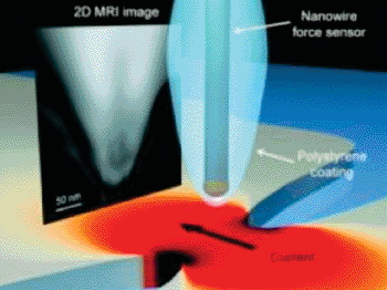
Image: Illustration of the experimental setup shows the two unique components of a novel MRI technique that was successful in producing a 2D MRI image with nanoscale spatial resolution (Photo courtesy of the University of Illinois).
Investigators have devised a unique nuclear magnetic resonance imaging (MRI) technique that delivers approximately 10-nm spatial resolutions.
This represents a significant advance in MRI sensitivity; current MRI techniques typically used in medical imaging generate spatial resolutions on the millimeter scale, with the highest-resolution research devices giving spatial resolution of a few micrometers.
“This is a very promising experimental result,” said the University of Illinois at Urbana-Champaign (U of I; USA) physicist Dr. Raffi Budakian, who led the research group. “Our approach brings MRI one step closer in its eventual progress toward atomic-scale imaging.”
MRI is used widely in clinical practice to differentiate pathologic tissue from normal tissue. It is noninvasive and harmless to the patient, using strong magnetic fields and nonionizing electromagnetic fields in the radiofrequency range, dissimilar to computed tomography (CT) scans and conventional X-rays, which both use more harmful ionizing radiation.
MRI uses static and time-dependent magnetic fields to detect the combined response of large ensembles of nuclear spins from molecules localized within millimeter-scale volumes in the body. Increasing the detection resolution from the millimeter to nanometer range would be a technologic dream come true.
The new technique introduces two novel components to overcome hurdles of applying traditional pulsed MR techniques in nanoscale systems. First, a unique protocol for spin manipulation applies periodic radiofrequency magnetic field pulses to encode temporal correlations in the statistical polarization of nuclear spins in the sample. Second, a nanoscale metal constriction focuses current, generating intense magnetic field-pulses.
In their proof-of-principal demonstration, the team used an ultrasensitive magnetic resonance sensor based on a silicon nanowire oscillator to reconstruct a two-dimensional projection image of the proton density in a polystyrene sample at nanoscale spatial resolution. “We expect this new technique to become a paradigm for nanoscale magnetic-resonance imaging and spectroscopy into the future,” added Dr. Budakian. “It is compatible with and can be incorporated into existing conventional MRI technologies.”
Related Links:
University of Illinois at Urbana-Champaign
Latest MRI News
- Simple Brain Scan Diagnoses Parkinson's Disease Years Before It Becomes Untreatable
- Cutting-Edge MRI Technology to Revolutionize Diagnosis of Common Heart Problem
- New MRI Technique Reveals True Heart Age to Prevent Attacks and Strokes
- AI Tool Predicts Relapse of Pediatric Brain Cancer from Brain MRI Scans
- AI Tool Tracks Effectiveness of Multiple Sclerosis Treatments Using Brain MRI Scans
- Ultra-Powerful MRI Scans Enable Life-Changing Surgery in Treatment-Resistant Epileptic Patients
- AI-Powered MRI Technology Improves Parkinson’s Diagnoses
- Biparametric MRI Combined with AI Enhances Detection of Clinically Significant Prostate Cancer
- First-Of-Its-Kind AI-Driven Brain Imaging Platform to Better Guide Stroke Treatment Options
- New Model Improves Comparison of MRIs Taken at Different Institutions
- Groundbreaking New Scanner Sees 'Previously Undetectable' Cancer Spread
- First-Of-Its-Kind Tool Analyzes MRI Scans to Measure Brain Aging
- AI-Enhanced MRI Images Make Cancerous Breast Tissue Glow
- AI Model Automatically Segments MRI Images
- New Research Supports Routine Brain MRI Screening in Asymptomatic Late-Stage Breast Cancer Patients
- Revolutionary Portable Device Performs Rapid MRI-Based Stroke Imaging at Patient's Bedside
Channels
Radiography
view channel
Machine Learning Algorithm Identifies Cardiovascular Risk from Routine Bone Density Scans
A new study published in the Journal of Bone and Mineral Research reveals that an automated machine learning program can predict the risk of cardiovascular events and falls or fractures by analyzing bone... Read more
AI Improves Early Detection of Interval Breast Cancers
Interval breast cancers, which occur between routine screenings, are easier to treat when detected earlier. Early detection can reduce the need for aggressive treatments and improve the chances of better outcomes.... Read more
World's Largest Class Single Crystal Diamond Radiation Detector Opens New Possibilities for Diagnostic Imaging
Diamonds possess ideal physical properties for radiation detection, such as exceptional thermal and chemical stability along with a quick response time. Made of carbon with an atomic number of six, diamonds... Read moreUltrasound
view channel
New Incision-Free Technique Halts Growth of Debilitating Brain Lesions
Cerebral cavernous malformations (CCMs), also known as cavernomas, are abnormal clusters of blood vessels that can grow in the brain, spinal cord, or other parts of the body. While most cases remain asymptomatic,... Read more.jpeg)
AI-Powered Lung Ultrasound Outperforms Human Experts in Tuberculosis Diagnosis
Despite global declines in tuberculosis (TB) rates in previous years, the incidence of TB rose by 4.6% from 2020 to 2023. Early screening and rapid diagnosis are essential elements of the World Health... Read moreNuclear Medicine
view channel
New Imaging Approach Could Reduce Need for Biopsies to Monitor Prostate Cancer
Prostate cancer is the second leading cause of cancer-related death among men in the United States. However, the majority of older men diagnosed with prostate cancer have slow-growing, low-risk forms of... Read more
Novel Radiolabeled Antibody Improves Diagnosis and Treatment of Solid Tumors
Interleukin-13 receptor α-2 (IL13Rα2) is a cell surface receptor commonly found in solid tumors such as glioblastoma, melanoma, and breast cancer. It is minimally expressed in normal tissues, making it... Read moreGeneral/Advanced Imaging
view channel
First-Of-Its-Kind Wearable Device Offers Revolutionary Alternative to CT Scans
Currently, patients with conditions such as heart failure, pneumonia, or respiratory distress often require multiple imaging procedures that are intermittent, disruptive, and involve high levels of radiation.... Read more
AI-Based CT Scan Analysis Predicts Early-Stage Kidney Damage Due to Cancer Treatments
Radioligand therapy, a form of targeted nuclear medicine, has recently gained attention for its potential in treating specific types of tumors. However, one of the potential side effects of this therapy... Read moreImaging IT
view channel
New Google Cloud Medical Imaging Suite Makes Imaging Healthcare Data More Accessible
Medical imaging is a critical tool used to diagnose patients, and there are billions of medical images scanned globally each year. Imaging data accounts for about 90% of all healthcare data1 and, until... Read more
Global AI in Medical Diagnostics Market to Be Driven by Demand for Image Recognition in Radiology
The global artificial intelligence (AI) in medical diagnostics market is expanding with early disease detection being one of its key applications and image recognition becoming a compelling consumer proposition... Read moreIndustry News
view channel
GE HealthCare and NVIDIA Collaboration to Reimagine Diagnostic Imaging
GE HealthCare (Chicago, IL, USA) has entered into a collaboration with NVIDIA (Santa Clara, CA, USA), expanding the existing relationship between the two companies to focus on pioneering innovation in... Read more
Patient-Specific 3D-Printed Phantoms Transform CT Imaging
New research has highlighted how anatomically precise, patient-specific 3D-printed phantoms are proving to be scalable, cost-effective, and efficient tools in the development of new CT scan algorithms... Read more
Siemens and Sectra Collaborate on Enhancing Radiology Workflows
Siemens Healthineers (Forchheim, Germany) and Sectra (Linköping, Sweden) have entered into a collaboration aimed at enhancing radiologists' diagnostic capabilities and, in turn, improving patient care... Read more


