fMRI Brain Scan Patterns Provide First Impartial Gauge of Pain
|
By MedImaging International staff writers Posted on 22 Apr 2013 |

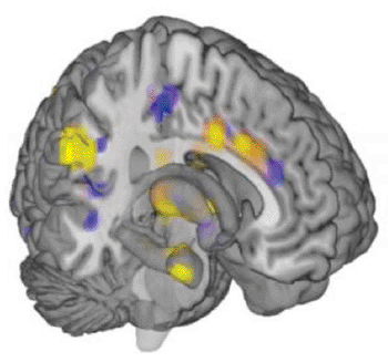
Image: Functional magnetic resonance imaging (fMRI) brain scan shows the neurologic signature for physical pain identified in a new study (Photo courtesy of Tor Wager, CU-Boulder).
Scientists for the first time have been able to forecast how much pain individuals are feeling by just looking at images of their brains.
The study, published on April 10, 2013, in the New England Journal of Medicine (NEJM), may lead to the development of effective ways that physicians can use to objectively quantify a patient’s pain. Currently, pain intensity can only be gauged based on a patient’s own description, which often includes rating the pain on a scale of one to 10. Objective measures of pain could confirm these pain reports and provide new insights into how the brain generates different types of pain.
The new findings also may provide the opportunity for the development of technology utilizing functional magnetic resonance imaging (fMRI) brain scans to objectively measure anger, anxiety, depression, or other emotional states. “Right now, there’s no clinically acceptable way to measure pain and other emotions other than to ask a person how they feel,” said Dr. Tor Wager, associate professor of psychology and neuroscience at University of Colorado Boulder (CU-Boulder; USA), and lead author of the paper.
The researchers, which included scientists from New York University (New York, NY, USA), Johns Hopkins University (Baltimore, MD, USA), and the University of Michigan, used computer data-mining techniques to sift through images of 114 brains that were captured when the study participants were exposed to multiple levels of heat, ranging from benignly warm to painfully hot. Utilizing the computer, the scientists identified a distinctive neurologic signature for the pain. “We found a pattern across multiple systems in the brain that is diagnostic of how much pain people feel in response to painful heat.” Dr. Wager said.
The researchers anticipated, conducting the research, that if a pain signature could be seen it would probably be distinctive to each individual. If that were the case, an individual’s pain level could only be predicted based on past images of his or her own brain. Instead, they found that the signature was transferable across different individuals, allowing the scientists to predict how much pain a person was being caused by the applied heat, with between 90%–100% accuracy, even with no earlier brain scans of that individual to use as a reference point.
The scientists also were startled to find that the signature was specific to physical pain. Earlier research has shown that social pain can appear very similar to physical pain in the way the brain activity it produces. One study, for example, revealed that the brain activity of people who have just been through a relationship breakup, and who were shown an image of the individual who rejected them, is similar to the brain activity of someone feeling physical pain.
However, when the investigators tested to see if the newly defined neurologic signature for heat pain would also pop up in the data collected earlier from the heartbroken participants, they found that the signature was absent. Ultimately, the scientists set out to see if the neurologic signature could detect when an analgesic was used to lessen the pain. The findings showed that the signature recorded a decrease in pain in subjects administered a painkiller.
The study’s findings revealed that the investigators cannot quantify physical pain, but they lay the potential for future research that could generate the first objective pain evaluations by clinicians and hospitals. To accomplish this, Dr. Wager and his colleagues are already examining how the neurologic signature can be verified when applied to different types of pain.
“I think there are many ways to extend this study, and we’re looking to test the patterns that we’ve developed for predicting pain across different conditions,” Dr. Wager said. “Is the predictive signature different if you experience pressure pain or mechanical pain, or pain on different parts of the body? We’re also looking towards using these same techniques to develop measures for chronic pain. The pattern we have found is not a measure of chronic pain, but we think it may be an ‘ingredient’ of chronic pain under some circumstances. Understanding the different contributions of different systems to chronic pain and other forms of suffering is an important step towards understanding and alleviating human suffering.”
Related Links:
University of Colorado Boulder
New York University
Johns Hopkins University
Latest MRI News
- New MRI Technique Reveals True Heart Age to Prevent Attacks and Strokes
- AI Tool Predicts Relapse of Pediatric Brain Cancer from Brain MRI Scans
- AI Tool Tracks Effectiveness of Multiple Sclerosis Treatments Using Brain MRI Scans
- Ultra-Powerful MRI Scans Enable Life-Changing Surgery in Treatment-Resistant Epileptic Patients
- AI-Powered MRI Technology Improves Parkinson’s Diagnoses
- Biparametric MRI Combined with AI Enhances Detection of Clinically Significant Prostate Cancer
- First-Of-Its-Kind AI-Driven Brain Imaging Platform to Better Guide Stroke Treatment Options
- New Model Improves Comparison of MRIs Taken at Different Institutions
- Groundbreaking New Scanner Sees 'Previously Undetectable' Cancer Spread
- First-Of-Its-Kind Tool Analyzes MRI Scans to Measure Brain Aging
- AI-Enhanced MRI Images Make Cancerous Breast Tissue Glow
- AI Model Automatically Segments MRI Images
- New Research Supports Routine Brain MRI Screening in Asymptomatic Late-Stage Breast Cancer Patients
- Revolutionary Portable Device Performs Rapid MRI-Based Stroke Imaging at Patient's Bedside
- AI Predicts After-Effects of Brain Tumor Surgery from MRI Scans
- MRI-First Strategy for Prostate Cancer Detection Proven Safe
Channels
Radiography
view channel
World's Largest Class Single Crystal Diamond Radiation Detector Opens New Possibilities for Diagnostic Imaging
Diamonds possess ideal physical properties for radiation detection, such as exceptional thermal and chemical stability along with a quick response time. Made of carbon with an atomic number of six, diamonds... Read more
AI-Powered Imaging Technique Shows Promise in Evaluating Patients for PCI
Percutaneous coronary intervention (PCI), also known as coronary angioplasty, is a minimally invasive procedure where small metal tubes called stents are inserted into partially blocked coronary arteries... Read moreUltrasound
view channel.jpeg)
AI-Powered Lung Ultrasound Outperforms Human Experts in Tuberculosis Diagnosis
Despite global declines in tuberculosis (TB) rates in previous years, the incidence of TB rose by 4.6% from 2020 to 2023. Early screening and rapid diagnosis are essential elements of the World Health... Read more
AI Identifies Heart Valve Disease from Common Imaging Test
Tricuspid regurgitation is a condition where the heart's tricuspid valve does not close completely during contraction, leading to backward blood flow, which can result in heart failure. A new artificial... Read moreNuclear Medicine
view channel
Novel Radiolabeled Antibody Improves Diagnosis and Treatment of Solid Tumors
Interleukin-13 receptor α-2 (IL13Rα2) is a cell surface receptor commonly found in solid tumors such as glioblastoma, melanoma, and breast cancer. It is minimally expressed in normal tissues, making it... Read more
Novel PET Imaging Approach Offers Never-Before-Seen View of Neuroinflammation
COX-2, an enzyme that plays a key role in brain inflammation, can be significantly upregulated by inflammatory stimuli and neuroexcitation. Researchers suggest that COX-2 density in the brain could serve... Read moreGeneral/Advanced Imaging
view channel
AI-Powered Imaging System Improves Lung Cancer Diagnosis
Given the need to detect lung cancer at earlier stages, there is an increasing need for a definitive diagnostic pathway for patients with suspicious pulmonary nodules. However, obtaining tissue samples... Read more
AI Model Significantly Enhances Low-Dose CT Capabilities
Lung cancer remains one of the most challenging diseases, making early diagnosis vital for effective treatment. Fortunately, advancements in artificial intelligence (AI) are revolutionizing lung cancer... Read moreImaging IT
view channel
New Google Cloud Medical Imaging Suite Makes Imaging Healthcare Data More Accessible
Medical imaging is a critical tool used to diagnose patients, and there are billions of medical images scanned globally each year. Imaging data accounts for about 90% of all healthcare data1 and, until... Read more
Global AI in Medical Diagnostics Market to Be Driven by Demand for Image Recognition in Radiology
The global artificial intelligence (AI) in medical diagnostics market is expanding with early disease detection being one of its key applications and image recognition becoming a compelling consumer proposition... Read moreIndustry News
view channel
GE HealthCare and NVIDIA Collaboration to Reimagine Diagnostic Imaging
GE HealthCare (Chicago, IL, USA) has entered into a collaboration with NVIDIA (Santa Clara, CA, USA), expanding the existing relationship between the two companies to focus on pioneering innovation in... Read more
Patient-Specific 3D-Printed Phantoms Transform CT Imaging
New research has highlighted how anatomically precise, patient-specific 3D-printed phantoms are proving to be scalable, cost-effective, and efficient tools in the development of new CT scan algorithms... Read more
Siemens and Sectra Collaborate on Enhancing Radiology Workflows
Siemens Healthineers (Forchheim, Germany) and Sectra (Linköping, Sweden) have entered into a collaboration aimed at enhancing radiologists' diagnostic capabilities and, in turn, improving patient care... Read more


