Consortium Established to Advance Integration of a Linear Accelerator with Simultaneous MRI Technology
|
By MedImaging International staff writers Posted on 06 Nov 2012 |
A consortium has been established to integrate radiotherapy delivery with simultaneous magnetic resonance imaging (MRI) to provide cutting-edge cancer treatments.
Elekta, AB (Stockholm, Sweden) and Philips Healthcare (Best, the Netherlands) reported that they will expand a joint program to develop new technology in cancer care with an imaging-treatment platform that merges radiation therapy and magnetic resonance imaging (MRI) technology in a single treatment system. The program for development will include a research consortium of leading radiation oncology centers and clinicians, which currently includes the University Medical Center Utrecht (the Netherlands).
The establishment of the consortium marks the next step in the collaboration between Elekta, a developer of neurosurgery and radiation therapy systems and Philips Healthcare, a leader in medical imaging systems. The consortium’s goal will be to fuse precise radiation delivery with MRI into one MRI-guided radiation therapy system. This will help clinicians to achieve unprecedented soft tissue imaging during radiation therapy and to adapt treatment delivery in real-time for very precise cancer treatments.
“Bringing the superior soft tissue imaging of MRI and precise radiotherapy together in one device could potentially revolutionize cancer care,” said Tomas Puusepp, president and CEO of Elekta. “The need to maximize therapeutic radiation on the target, while minimizing the exposure of healthy tissue is entirely driven by the best interests of the patient--they deserve the best chance for a cure and an improved quality of life. Elekta and Philips are leaders in the global healthcare community with a complete spectrum of expertise to fulfill this vision.”
“Cancer is a major global disease that we hope to control with more targeted treatments,” remarked Gene Saragnese, CEO imaging systems at Philips Healthcare. “MRI is emerging in oncology applications because of its excellent real-time 3D [three-dimensional] visualization of soft tissue. Together with our partners, all leaders in radiation therapy delivery, we are convinced that the integrated MRI-guided radiation therapy system has the potential to become a game changer in cancer care on a global scale.”
Medical device companies have constructed and evaluated a prototype system, working with University Medical Center Utrecht, that incorporates a linear accelerator and a 1.5-Tesla MRI system. The promising findings of preliminary testing has enabled the project to take the effort to the next level of development and testing by a select group of consortium partners.
“We are proud and excited about this project,” commented Prof. Jan Lagendijk, department of radiotherapy, University Medical Center Utrecht. “Elekta, Philips, and my department have strived for over a decade to make this possible. Through real-time imaging of both tumors and organs at risk, an integrated MRI-guided radiation therapy system would enable us to see more precisely than ever what we treat, and irradiate just the tumor as it moves in the body during treatment. This could potentially bring significant benefits to patients and overall healthcare economics.”
Radiotherapy is one of the standard tools utilized to treat cancer, either as a treatment plan by itself or used with other modalities such as chemotherapy. The technique involves targeting cancerous tissue and irradiating it with high-energy radiation beams in a manner that maximizes sparing of healthy tissue near the tumor. Radiation therapy delivered by linear accelerators and medical imaging already plays a fundamental role in treatment planning, delivery, and postcare, and is a cost-effective and safe way to treat patients with cancer.
The integrated MRI-guided radiation therapy system is currently in development and not available for sale.
Elekta develops applications and treatment planning systems for radiotherapy, radiosurgery, and brachytherapy, as well as workflow enhancing software systems across the spectrum of cancer care. Elekta oncology and neurosurgery systems are used in over 6,000 hospitals worldwide. Elekta employs approximately 3,400 employees worldwide.
Related Links:
Elekta
Philips Healthcare
Latest Industry News News
- GE HealthCare and NVIDIA Collaboration to Reimagine Diagnostic Imaging
- Patient-Specific 3D-Printed Phantoms Transform CT Imaging
- Siemens and Sectra Collaborate on Enhancing Radiology Workflows
- Bracco Diagnostics and ColoWatch Partner to Expand Availability CRC Screening Tests Using Virtual Colonoscopy
- Mindray Partners with TeleRay to Streamline Ultrasound Delivery
- Philips and Medtronic Partner on Stroke Care
- Siemens and Medtronic Enter into Global Partnership for Advancing Spine Care Imaging Technologies
- RSNA 2024 Technical Exhibits to Showcase Latest Advances in Radiology
- Bracco Collaborates with Arrayus on Microbubble-Assisted Focused Ultrasound Therapy for Pancreatic Cancer
- Innovative Collaboration to Enhance Ischemic Stroke Detection and Elevate Standards in Diagnostic Imaging
- RSNA 2024 Registration Opens
- Microsoft collaborates with Leading Academic Medical Systems to Advance AI in Medical Imaging
- GE HealthCare Acquires Intelligent Ultrasound Group’s Clinical Artificial Intelligence Business
- Bayer and Rad AI Collaborate on Expanding Use of Cutting Edge AI Radiology Operational Solutions
- Polish Med-Tech Company BrainScan to Expand Extensively into Foreign Markets
- Hologic Acquires UK-Based Breast Surgical Guidance Company Endomagnetics Ltd.
Channels
Radiography
view channel
AI Improves Early Detection of Interval Breast Cancers
Interval breast cancers, which occur between routine screenings, are easier to treat when detected earlier. Early detection can reduce the need for aggressive treatments and improve the chances of better outcomes.... Read more
World's Largest Class Single Crystal Diamond Radiation Detector Opens New Possibilities for Diagnostic Imaging
Diamonds possess ideal physical properties for radiation detection, such as exceptional thermal and chemical stability along with a quick response time. Made of carbon with an atomic number of six, diamonds... Read moreMRI
view channel
Cutting-Edge MRI Technology to Revolutionize Diagnosis of Common Heart Problem
Aortic stenosis is a common and potentially life-threatening heart condition. It occurs when the aortic valve, which regulates blood flow from the heart to the rest of the body, becomes stiff and narrow.... Read more
New MRI Technique Reveals True Heart Age to Prevent Attacks and Strokes
Heart disease remains one of the leading causes of death worldwide. Individuals with conditions such as diabetes or obesity often experience accelerated aging of their hearts, sometimes by decades.... Read more
AI Tool Predicts Relapse of Pediatric Brain Cancer from Brain MRI Scans
Many pediatric gliomas are treatable with surgery alone, but relapses can be catastrophic. Predicting which patients are at risk for recurrence remains challenging, leading to frequent follow-ups with... Read more
AI Tool Tracks Effectiveness of Multiple Sclerosis Treatments Using Brain MRI Scans
Multiple sclerosis (MS) is a condition in which the immune system attacks the brain and spinal cord, leading to impairments in movement, sensation, and cognition. Magnetic Resonance Imaging (MRI) markers... Read moreUltrasound
view channel.jpeg)
AI-Powered Lung Ultrasound Outperforms Human Experts in Tuberculosis Diagnosis
Despite global declines in tuberculosis (TB) rates in previous years, the incidence of TB rose by 4.6% from 2020 to 2023. Early screening and rapid diagnosis are essential elements of the World Health... Read more
AI Identifies Heart Valve Disease from Common Imaging Test
Tricuspid regurgitation is a condition where the heart's tricuspid valve does not close completely during contraction, leading to backward blood flow, which can result in heart failure. A new artificial... Read moreNuclear Medicine
view channel
Novel Radiolabeled Antibody Improves Diagnosis and Treatment of Solid Tumors
Interleukin-13 receptor α-2 (IL13Rα2) is a cell surface receptor commonly found in solid tumors such as glioblastoma, melanoma, and breast cancer. It is minimally expressed in normal tissues, making it... Read more
Novel PET Imaging Approach Offers Never-Before-Seen View of Neuroinflammation
COX-2, an enzyme that plays a key role in brain inflammation, can be significantly upregulated by inflammatory stimuli and neuroexcitation. Researchers suggest that COX-2 density in the brain could serve... Read moreGeneral/Advanced Imaging
view channel
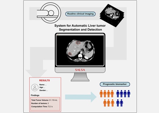
CT-Based Deep Learning-Driven Tool to Enhance Liver Cancer Diagnosis
Medical imaging, such as computed tomography (CT) scans, plays a crucial role in oncology, offering essential data for cancer detection, treatment planning, and monitoring of response to therapies.... Read more
AI-Powered Imaging System Improves Lung Cancer Diagnosis
Given the need to detect lung cancer at earlier stages, there is an increasing need for a definitive diagnostic pathway for patients with suspicious pulmonary nodules. However, obtaining tissue samples... Read moreImaging IT
view channel
New Google Cloud Medical Imaging Suite Makes Imaging Healthcare Data More Accessible
Medical imaging is a critical tool used to diagnose patients, and there are billions of medical images scanned globally each year. Imaging data accounts for about 90% of all healthcare data1 and, until... Read more


