Traumatic Brain Injury Identified with High-Definition Fiber Tracking
|
By MedImaging International staff writers Posted on 15 Mar 2012 |

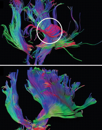
Image: Diffusion tensor imaging fiber tracking (upper) and HDFT (lower) in the TBI patient at 17 weeks post-injury. The DTI fiber tracking shows inaccurate fiber directions and termination points (white circle), whereas the HDFT scan shows accurate details of damaged areas of the right anterior corona radiata without false turns or false continuations (Photo courtesy of the University of Pittsburgh).
An effective new imaging modality called high-definition fiber tracking (HDFT) will allow clinicians to precisely see for the first time neural connections shattered by traumatic brain injury (TBI) and other neurologic disorders, similar to X-rays showing a fractured bone.
The researchers, from the University of Pittsburgh (UPitt; PA, USA), published their findings in a report published online March 3, 2012, in the Journal of Neurosurgery. In the report, the researchers described the instance of a 32-year-old man who was not wearing a helmet when his all-terrain vehicle crashed. At first, his computed tomography (CT) scans showed bleeding and swelling on the right side of the brain, the side that controls left-sided body movement. One week later, while the man was still in a coma, a traditional magnetic resonance imaging (MRI) scan revealed brain bruising and swelling in the same area. When he awoke three weeks later, the man could not move his left leg, arm, and hand.
“There are about 1.7 million cases of TBI in the country each year, and all too often conventional scans show no injury or show improvement over time even though the patient continues to struggle,” said cosenior author and UPMC neurosurgeon David O. Okonkwo, MD, PhD, associate professor, department of neurological surgery, Pitt School of Medicine. "Until now, we have had no objective way of identifying how the injury damaged the patient’s brain tissue, predicting how the patient would fare, or planning rehabilitation to maximize the recovery.”
HDFT might be able to provide those answers, said cosenior author Walter Schneider, PhD, professor of psychology at Pitt’s Learning Research and Development Center (LRDC), who led the team that developed the technology. Information from advanced MRI scanners is processed through computer algorithms to reveal the wiring of the brain in remarkable detail and to pinpoint breaks in the cables, called fiber tracts. Each tract contains millions of neuronal connections.
“In our experiments, HDFT has been able to identify disruptions in neural pathways with a clarity that no other method can see,” Dr. Schneider said. “With it, we can virtually dissect 40 major fiber tracts in the brain to find damaged areas and quantify the proportion of fibers lost relative to the uninjured side of the brain or to the brains of healthy individuals. Now, we can clearly see breaks and identify which parts of the brain have lost connections.”
HDFT scans of the study patient’s brain were performed 4 and 10 months after he was injured; he also had another scan performed with current state-of the-art diffusion tensor imaging (DTI), an imaging modality that collects data points from 51 directions, while HDFT is based on data from 257 directions. For the latter, the injury site was compared to the healthy side of his brain, as well as to HDFT brain scans from six healthy individuals.
Only the HDFT scan detected a lesion in a motor fiber pathway of the brain that correlated with the patient’s symptoms of left-sided weakness, including mostly intact fibers in the area controlling his left leg and extensive breaks in the region controlling his left hand. The patient ultimately recovered movement in his left leg and arm by six months after the accident, but still could not use his wrist and fingers effectively 10 months later.
Memory loss, language difficulties, personality changes, and other brain alterations occur with TBI, which the researchers are exploring with HDFT in other research protocols.
UPMC neurosurgeons also have utilized the imaging technique to supplement conventional imaging, noted Robert Friedlander, MD, professor and chair, department of neurological surgery, Pitt School of Medicine, and UPitt Medical Center (UPMC) professor of neurosurgery and neurobiology. “I have used HDFT scans to map my approach to removing certain tumors and vascular abnormalities that lie in areas of the brain that cannot be reached without going through normal tissue,” he said. “It shows me where significant functional pathways are relative to the lesion, so that I can make better decisions about which fiber tracts must be avoided and what might be an acceptable sacrifice to maintain the patient’s best quality of life after surgery.”
Dr. Okonkwo noted that the patient and his family were reassured to learn that there was evidence of brain damage to clarify his ongoing difficulties. The researchers continue to assess and validate HDFT’s usefulness as a brain-imaging tool, so it is not yet routinely available. “We have been wowed by the detailed, meaningful images we can get with this technology,” Dr. Okonkwo noted. “HDFT has the potential to be a game-changer in the way we handle TBI and other brain disorders.”
Related Links:
University of Pittsburgh
Latest MRI News
- AI Tool Tracks Effectiveness of Multiple Sclerosis Treatments Using Brain MRI Scans
- Ultra-Powerful MRI Scans Enable Life-Changing Surgery in Treatment-Resistant Epileptic Patients
- AI-Powered MRI Technology Improves Parkinson’s Diagnoses
- Biparametric MRI Combined with AI Enhances Detection of Clinically Significant Prostate Cancer
- First-Of-Its-Kind AI-Driven Brain Imaging Platform to Better Guide Stroke Treatment Options
- New Model Improves Comparison of MRIs Taken at Different Institutions
- Groundbreaking New Scanner Sees 'Previously Undetectable' Cancer Spread
- First-Of-Its-Kind Tool Analyzes MRI Scans to Measure Brain Aging
- AI-Enhanced MRI Images Make Cancerous Breast Tissue Glow
- AI Model Automatically Segments MRI Images
- New Research Supports Routine Brain MRI Screening in Asymptomatic Late-Stage Breast Cancer Patients
- Revolutionary Portable Device Performs Rapid MRI-Based Stroke Imaging at Patient's Bedside
- AI Predicts After-Effects of Brain Tumor Surgery from MRI Scans
- MRI-First Strategy for Prostate Cancer Detection Proven Safe
- First-Of-Its-Kind 10' x 48' Mobile MRI Scanner Transforms User and Patient Experience
- New Model Makes MRI More Accurate and Reliable
Channels
Radiography
view channel
World's Largest Class Single Crystal Diamond Radiation Detector Opens New Possibilities for Diagnostic Imaging
Diamonds possess ideal physical properties for radiation detection, such as exceptional thermal and chemical stability along with a quick response time. Made of carbon with an atomic number of six, diamonds... Read more
AI-Powered Imaging Technique Shows Promise in Evaluating Patients for PCI
Percutaneous coronary intervention (PCI), also known as coronary angioplasty, is a minimally invasive procedure where small metal tubes called stents are inserted into partially blocked coronary arteries... Read moreUltrasound
view channel.jpeg)
AI-Powered Lung Ultrasound Outperforms Human Experts in Tuberculosis Diagnosis
Despite global declines in tuberculosis (TB) rates in previous years, the incidence of TB rose by 4.6% from 2020 to 2023. Early screening and rapid diagnosis are essential elements of the World Health... Read more
AI Identifies Heart Valve Disease from Common Imaging Test
Tricuspid regurgitation is a condition where the heart's tricuspid valve does not close completely during contraction, leading to backward blood flow, which can result in heart failure. A new artificial... Read moreNuclear Medicine
view channel
Novel Radiolabeled Antibody Improves Diagnosis and Treatment of Solid Tumors
Interleukin-13 receptor α-2 (IL13Rα2) is a cell surface receptor commonly found in solid tumors such as glioblastoma, melanoma, and breast cancer. It is minimally expressed in normal tissues, making it... Read more
Novel PET Imaging Approach Offers Never-Before-Seen View of Neuroinflammation
COX-2, an enzyme that plays a key role in brain inflammation, can be significantly upregulated by inflammatory stimuli and neuroexcitation. Researchers suggest that COX-2 density in the brain could serve... Read moreGeneral/Advanced Imaging
view channel
AI-Powered Imaging System Improves Lung Cancer Diagnosis
Given the need to detect lung cancer at earlier stages, there is an increasing need for a definitive diagnostic pathway for patients with suspicious pulmonary nodules. However, obtaining tissue samples... Read more
AI Model Significantly Enhances Low-Dose CT Capabilities
Lung cancer remains one of the most challenging diseases, making early diagnosis vital for effective treatment. Fortunately, advancements in artificial intelligence (AI) are revolutionizing lung cancer... Read moreImaging IT
view channel
New Google Cloud Medical Imaging Suite Makes Imaging Healthcare Data More Accessible
Medical imaging is a critical tool used to diagnose patients, and there are billions of medical images scanned globally each year. Imaging data accounts for about 90% of all healthcare data1 and, until... Read more
Global AI in Medical Diagnostics Market to Be Driven by Demand for Image Recognition in Radiology
The global artificial intelligence (AI) in medical diagnostics market is expanding with early disease detection being one of its key applications and image recognition becoming a compelling consumer proposition... Read moreIndustry News
view channel
GE HealthCare and NVIDIA Collaboration to Reimagine Diagnostic Imaging
GE HealthCare (Chicago, IL, USA) has entered into a collaboration with NVIDIA (Santa Clara, CA, USA), expanding the existing relationship between the two companies to focus on pioneering innovation in... Read more
Patient-Specific 3D-Printed Phantoms Transform CT Imaging
New research has highlighted how anatomically precise, patient-specific 3D-printed phantoms are proving to be scalable, cost-effective, and efficient tools in the development of new CT scan algorithms... Read more
Siemens and Sectra Collaborate on Enhancing Radiology Workflows
Siemens Healthineers (Forchheim, Germany) and Sectra (Linköping, Sweden) have entered into a collaboration aimed at enhancing radiologists' diagnostic capabilities and, in turn, improving patient care... Read more


