TOF PET/MR Scanner to Advance New Approaches to Cancer and Neurology
|
By MedImaging International staff writers Posted on 24 Jan 2012 |

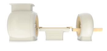
Image: The Ingenuity TF PET/MR scanner (Photo courtesy of Philips Healthcare).
A university medical center in Amsterdam will be the first hospital in the Netherlands to have sophisticated time-of-flight (TOF) positron emission tomography/magnetic resonance (PET/MR) scanner technology.
Philips Healthcare (Best, The Netherlands) and the VU University Medical Center Amsterdam (The Netherlands) announced that they have signed an agreement to install one of Philips’ highly innovative Ingenuity TF PET/MR scanners at the center. This advanced imaging technology will help researchers at the VU University Medical Center Amsterdam to investigate new methods of diagnosing and treating cancer and neurologic disorders.
The decision to install the scanner was fueled by the need for diagnostic solutions that allow clinicians to bring customized healthcare to their patients. The VU University Medical Center Amsterdam is a globally recognized center of excellence in the fields of oncology, neurology, and cardiology. It has specific expertise in imaging technologies, such as PET, that use targeted radioactive tracers to produce three-dimensional (3D) images of organs such as the brain or internal lesions such as tumors.
“The VU University Medical Center Amsterdam is particularly strong in the development and clinical application of PET technology in the fields of oncology, neurology, and cardiology,” said Wim Stalman, dean and vice chairman of the board of the VU University Medical Center Amsterdam. “It is therefore particularly exciting to work with Philips, a true innovator in the field. I am convinced that together we will bring hybrid PET technologies such as PET/MR to the next level.”
“Constant innovation in medical imaging technologies has significantly expanded the frontiers of modern healthcare,” said Richard Fabian, general manager nuclear medicine, Philips Healthcare. “Cancer care is an innovation focus area for Philips, in which new imaging modalities such as Philips’ PET/MR system are expected to play an ever-increasing role in the diagnosis, treatment and monitoring of the disease. We look forward to our continuing collaboration with the VU University Medical Center Amsterdam to fully exploit the potential benefits of PET/MR imaging in patient care.”
Philips’ Ingenuity TF PET/MR system is one of the new imaging solutions that the company has recently released under the banner Imaging 2.0, an initiative developed to address the needs of radiologists and to advance clinical excellence through greater collaboration and integration combined with increased patient focus and enhanced cost-effectiveness.
The Ingenuity TF PET/MR scanner combines the molecular imaging capabilities of PET with the excellent soft tissue imaging capabilities of MR imaging. PET and MR have been used as separate and distinctive imaging modalities for several years, with each modality requiring its own suite of rooms to house the necessary equipment. Philips was the first company effectively to overcome the technical challenges involved in bringing these two imaging technologies into close physical proximity in a whole-body scanner, so that sequential PET and MR images can be acquired in the same session. This allows very accurate overlaying of the PET and MR images so that clinicians can combine the functional and anatomic data provided by PET and MR, respectively, into one fused image.
Philips has a long-standing relationship with the VU University Medical Center Amsterdam in leading-edge medical research--for example, via research organizations such as the Netherlands’ Center for Translational Molecular Medicine (CTMM).
Related Links:
Philips Healthcare
VU University Medical Center Amsterdam
Latest Nuclear Medicine News
- Novel PET Imaging Approach Offers Never-Before-Seen View of Neuroinflammation
- Novel Radiotracer Identifies Biomarker for Triple-Negative Breast Cancer
- Innovative PET Imaging Technique to Help Diagnose Neurodegeneration
- New Molecular Imaging Test to Improve Lung Cancer Diagnosis
- Novel PET Technique Visualizes Spinal Cord Injuries to Predict Recovery
- Next-Gen Tau Radiotracers Outperform FDA-Approved Imaging Agents in Detecting Alzheimer’s
- Breakthrough Method Detects Inflammation in Body Using PET Imaging
- Advanced Imaging Reveals Hidden Metastases in High-Risk Prostate Cancer Patients
- Combining Advanced Imaging Technologies Offers Breakthrough in Glioblastoma Treatment
- New Molecular Imaging Agent Accurately Identifies Crucial Cancer Biomarker
- New Scans Light Up Aggressive Tumors for Better Treatment
- AI Stroke Brain Scan Readings Twice as Accurate as Current Method
- AI Analysis of PET/CT Images Predicts Side Effects of Immunotherapy in Lung Cancer
- New Imaging Agent to Drive Step-Change for Brain Cancer Imaging
- Portable PET Scanner to Detect Earliest Stages of Alzheimer’s Disease
- New Immuno-PET Imaging Technique Identifies Glioblastoma Patients Who Would Benefit from Immunotherapy
Channels
Radiography
view channel
World's Largest Class Single Crystal Diamond Radiation Detector Opens New Possibilities for Diagnostic Imaging
Diamonds possess ideal physical properties for radiation detection, such as exceptional thermal and chemical stability along with a quick response time. Made of carbon with an atomic number of six, diamonds... Read more
AI-Powered Imaging Technique Shows Promise in Evaluating Patients for PCI
Percutaneous coronary intervention (PCI), also known as coronary angioplasty, is a minimally invasive procedure where small metal tubes called stents are inserted into partially blocked coronary arteries... Read moreMRI
view channel
AI Tool Tracks Effectiveness of Multiple Sclerosis Treatments Using Brain MRI Scans
Multiple sclerosis (MS) is a condition in which the immune system attacks the brain and spinal cord, leading to impairments in movement, sensation, and cognition. Magnetic Resonance Imaging (MRI) markers... Read more
Ultra-Powerful MRI Scans Enable Life-Changing Surgery in Treatment-Resistant Epileptic Patients
Approximately 360,000 individuals in the UK suffer from focal epilepsy, a condition in which seizures spread from one part of the brain. Around a third of these patients experience persistent seizures... Read more
AI-Powered MRI Technology Improves Parkinson’s Diagnoses
Current research shows that the accuracy of diagnosing Parkinson’s disease typically ranges from 55% to 78% within the first five years of assessment. This is partly due to the similarities shared by Parkinson’s... Read more
Biparametric MRI Combined with AI Enhances Detection of Clinically Significant Prostate Cancer
Artificial intelligence (AI) technologies are transforming the way medical images are analyzed, offering unprecedented capabilities in quantitatively extracting features that go beyond traditional visual... Read moreUltrasound
view channel
AI Identifies Heart Valve Disease from Common Imaging Test
Tricuspid regurgitation is a condition where the heart's tricuspid valve does not close completely during contraction, leading to backward blood flow, which can result in heart failure. A new artificial... Read more
Novel Imaging Method Enables Early Diagnosis and Treatment Monitoring of Type 2 Diabetes
Type 2 diabetes is recognized as an autoimmune inflammatory disease, where chronic inflammation leads to alterations in pancreatic islet microvasculature, a key factor in β-cell dysfunction.... Read moreGeneral/Advanced Imaging
view channel
AI-Powered Imaging System Improves Lung Cancer Diagnosis
Given the need to detect lung cancer at earlier stages, there is an increasing need for a definitive diagnostic pathway for patients with suspicious pulmonary nodules. However, obtaining tissue samples... Read more
AI Model Significantly Enhances Low-Dose CT Capabilities
Lung cancer remains one of the most challenging diseases, making early diagnosis vital for effective treatment. Fortunately, advancements in artificial intelligence (AI) are revolutionizing lung cancer... Read moreImaging IT
view channel
New Google Cloud Medical Imaging Suite Makes Imaging Healthcare Data More Accessible
Medical imaging is a critical tool used to diagnose patients, and there are billions of medical images scanned globally each year. Imaging data accounts for about 90% of all healthcare data1 and, until... Read more
Global AI in Medical Diagnostics Market to Be Driven by Demand for Image Recognition in Radiology
The global artificial intelligence (AI) in medical diagnostics market is expanding with early disease detection being one of its key applications and image recognition becoming a compelling consumer proposition... Read moreIndustry News
view channel
GE HealthCare and NVIDIA Collaboration to Reimagine Diagnostic Imaging
GE HealthCare (Chicago, IL, USA) has entered into a collaboration with NVIDIA (Santa Clara, CA, USA), expanding the existing relationship between the two companies to focus on pioneering innovation in... Read more
Patient-Specific 3D-Printed Phantoms Transform CT Imaging
New research has highlighted how anatomically precise, patient-specific 3D-printed phantoms are proving to be scalable, cost-effective, and efficient tools in the development of new CT scan algorithms... Read more
Siemens and Sectra Collaborate on Enhancing Radiology Workflows
Siemens Healthineers (Forchheim, Germany) and Sectra (Linköping, Sweden) have entered into a collaboration aimed at enhancing radiologists' diagnostic capabilities and, in turn, improving patient care... Read more


