Photon-Counting CT Beats Dual-Source CT at Detecting Heart Defects in Babies
|
By MedImaging International staff writers Posted on 25 May 2023 |

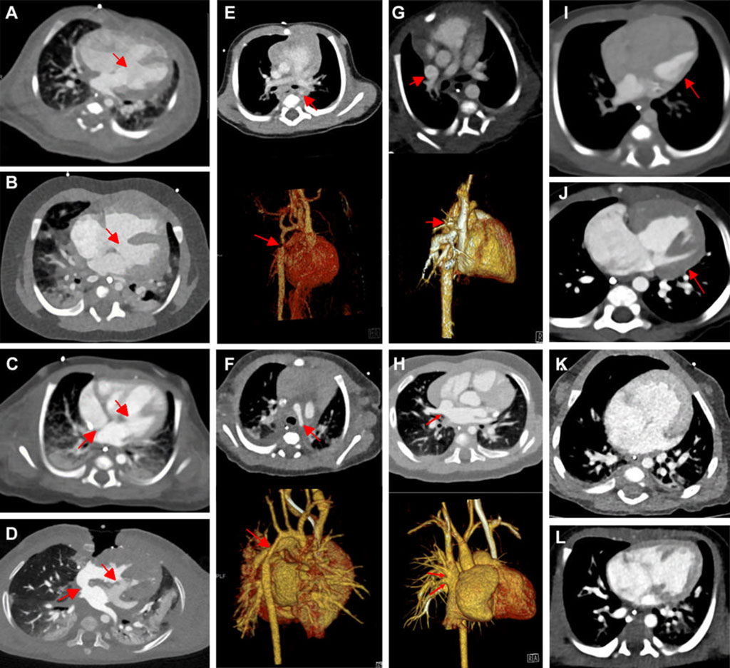
In the neonatal stage, congenital heart defects emerge as the leading cause of sickness and death, impacting nearly one percent of all live births. Around a quarter of these defects are severe, necessitating surgical treatments within the first month post-birth. In order to plan for the operation and devise virtual and printed 3D heart models, extensive evaluation using ultrasound, MRI, and CT scans is usually required. Now, a study has found that a new advanced form of CT imaging called photon-counting computed tomography (PCCT) offers superior cardiovascular imaging quality at a comparable radiation dose to dual-source CT (DSCT) for infants with suspected cardiac defects.
PCCT is an emerging imaging technique that records the exact count and energy measurements of incoming X-ray photons. In comparison to DSCT, PCCT delivers higher image resolution and/or lower radiation doses, a feature that is especially useful in pediatric imaging. Although the efficacy of PCCT in enhancing cardiovascular CT imaging in adults has been established, there is a dearth of information about its application in neonates and young children. For their study, researchers at RWTH Aachen University Hospital (Aachen, Germany) assessed pre-existing clinical CT scans of 113 children who had undergone contrast-enhanced PCCT (30 infants), DSCT (83 infants), or both (one infant) for their heart and thoracic aorta from January 2019 through October 2022. The group under study was comprised of 55 girls and 58 boys, with a median age of 66 days.
The researchers found that the PCCT images were sharper, had less image noise, and demonstrated higher contrast than DSCT images. The mean overall visual quality of the images was higher for PCCT than DSCT, using a similar radiation dose. Over 97% of PCCT images met the diagnostic quality criteria, as opposed to 77% of DSCT images. It was also observed by the research team that almost one-fourth of the DSCT images were of restricted or non-diagnostic quality, and 40% were of moderate quality.
“In our study, none of the PCCT examinations exhibited a poor image quality, and only a few were of limited or moderate quality,” said Timm Dirrichs, M.D., senior physician and specialist in cardiothoracic radiology in the Department of Diagnostic and Interventional Radiology at RWTH Aachen University Hospital. “PCCT is a promising method that may improve diagnostic image quality and efficiency compared to DSCT imaging. This higher efficiency can be used to reduce the radiation dose at a given image quality level or to improve image quality at a given radiation level.”
Related Links:
RWTH Aachen University Hospital
Latest General/Advanced Imaging News
- Bone Density Test Uses Existing CT Images to Predict Fractures
- AI Predicts Cardiac Risk and Mortality from Routine Chest CT Scans
- Radiation Therapy Computed Tomography Solution Boosts Imaging Accuracy
- PET Scans Reveal Hidden Inflammation in Multiple Sclerosis Patients
- Artificial Intelligence Evaluates Cardiovascular Risk from CT Scans
- New AI Method Captures Uncertainty in Medical Images
- CT Coronary Angiography Reduces Need for Invasive Tests to Diagnose Coronary Artery Disease
- Novel Blood Test Could Reduce Need for PET Imaging of Patients with Alzheimer’s
- CT-Based Deep Learning Algorithm Accurately Differentiates Benign From Malignant Vertebral Fractures
- Minimally Invasive Procedure Could Help Patients Avoid Thyroid Surgery
- Self-Driving Mobile C-Arm Reduces Imaging Time during Surgery
- AR Application Turns Medical Scans Into Holograms for Assistance in Surgical Planning
- Imaging Technology Provides Ground-Breaking New Approach for Diagnosing and Treating Bowel Cancer
- CT Coronary Calcium Scoring Predicts Heart Attacks and Strokes
- AI Model Detects 90% of Lymphatic Cancer Cases from PET and CT Images
- Breakthrough Technology Revolutionizes Breast Imaging
Channels
Radiography
view channel
Novel Breast Imaging System Proves As Effective As Mammography
Breast cancer remains the most frequently diagnosed cancer among women. It is projected that one in eight women will be diagnosed with breast cancer during her lifetime, and one in 42 women who turn 50... Read more
AI Assistance Improves Breast-Cancer Screening by Reducing False Positives
Radiologists typically detect one case of cancer for every 200 mammograms reviewed. However, these evaluations often result in false positives, leading to unnecessary patient recalls for additional testing,... Read moreMRI
view channel
Low-Cost Whole-Body MRI Device Combined with AI Generates High-Quality Results
Magnetic Resonance Imaging (MRI) has significantly transformed healthcare, providing a noninvasive, radiation-free method for detailed imaging. It is especially promising for the future of medical diagnosis... Read more
World's First Whole-Body Ultra-High Field MRI Officially Comes To Market
The world's first whole-body ultra-high field (UHF) MRI has officially come to market, marking a remarkable advancement in diagnostic radiology. United Imaging (Shanghai, China) has secured clearance from the U.... Read moreUltrasound
view channel.jpg)
Diagnostic System Automatically Analyzes TTE Images to Identify Congenital Heart Disease
Congenital heart disease (CHD) is one of the most prevalent congenital anomalies worldwide, presenting substantial health and financial challenges for affected patients. Early detection and treatment of... Read more
Super-Resolution Imaging Technique Could Improve Evaluation of Cardiac Conditions
The heart depends on efficient blood circulation to pump blood throughout the body, delivering oxygen to tissues and removing carbon dioxide and waste. Yet, when heart vessels are damaged, it can disrupt... Read more
First AI-Powered POC Ultrasound Diagnostic Solution Helps Prioritize Cases Based On Severity
Ultrasound scans are essential for identifying and diagnosing various medical conditions, but often, patients must wait weeks or months for results due to a shortage of qualified medical professionals... Read moreNuclear Medicine
view channel
New PET Biomarker Predicts Success of Immune Checkpoint Blockade Therapy
Immunotherapies, such as immune checkpoint blockade (ICB), have shown promising clinical results in treating melanoma, non-small cell lung cancer, and other tumor types. However, the effectiveness of these... Read moreNew PET Agent Rapidly and Accurately Visualizes Lesions in Clear Cell Renal Cell Carcinoma Patients
Clear cell renal cell cancer (ccRCC) represents 70-80% of renal cell carcinoma cases. While localized disease can be effectively treated with surgery and ablative therapies, one-third of patients either... Read more
New Imaging Technique Monitors Inflammation Disorders without Radiation Exposure
Imaging inflammation using traditional radiological techniques presents significant challenges, including radiation exposure, poor image quality, high costs, and invasive procedures. Now, new contrast... Read more
New SPECT/CT Technique Could Change Imaging Practices and Increase Patient Access
The development of lead-212 (212Pb)-PSMA–based targeted alpha therapy (TAT) is garnering significant interest in treating patients with metastatic castration-resistant prostate cancer. The imaging of 212Pb,... Read moreImaging IT
view channel
New Google Cloud Medical Imaging Suite Makes Imaging Healthcare Data More Accessible
Medical imaging is a critical tool used to diagnose patients, and there are billions of medical images scanned globally each year. Imaging data accounts for about 90% of all healthcare data1 and, until... Read more
Global AI in Medical Diagnostics Market to Be Driven by Demand for Image Recognition in Radiology
The global artificial intelligence (AI) in medical diagnostics market is expanding with early disease detection being one of its key applications and image recognition becoming a compelling consumer proposition... Read moreIndustry News
view channel
Hologic Acquires UK-Based Breast Surgical Guidance Company Endomagnetics Ltd.
Hologic, Inc. (Marlborough, MA, USA) has entered into a definitive agreement to acquire Endomagnetics Ltd. (Cambridge, UK), a privately held developer of breast cancer surgery technologies, for approximately... Read more
Bayer and Google Partner on New AI Product for Radiologists
Medical imaging data comprises around 90% of all healthcare data, and it is a highly complex and rich clinical data modality and serves as a vital tool for diagnosing patients. Each year, billions of medical... Read more


