CADx System Combines AI with Low-Cost MRI Scans to Speed up Prostate Cancer Detection
|
By MedImaging International staff writers Posted on 29 Mar 2023 |

The current protocol for treating prostate cancer involves a number of uncomfortable and unreliable procedures. One example is the PSA test, which measures the level of prostate-specific antigen (PSA) in the blood and can sometimes indicate the presence of prostate cancer. However, this test is not conclusive and often leads to additional testing such as MRI to identify any abnormalities in the prostate. The development of new blood, urine or tissue tests for prostate cancer could be very promising but such tests do not reveal the location or extent of the cancer, which is something that imaging with artificial intelligence (AI) can do.
Similarly, digital rectal exams (DREs) have been found to be just slightly over 50% accurate due to the significant number of prostate cancers located in the anterior aspect of the gland, which is far from the rectum and cannot be felt. In addition, while biopsies can confirm the presence of cancer, their random or targeted sampling often miss cancerous lesions. Biopsies are also costly, carry a risk of infection, and can lead to false positives. Therefore, biopsies are not an effective screening method for prostate cancer but can confirm a diagnosis if used in conjunction with imaging guidance. Now, a powerful computer-aided diagnosis (CADx) software device is poised to change the standard of care for prostate cancer screening, detection and diagnosis by avoiding the expensive pitfalls of current prostate cancer pathways.
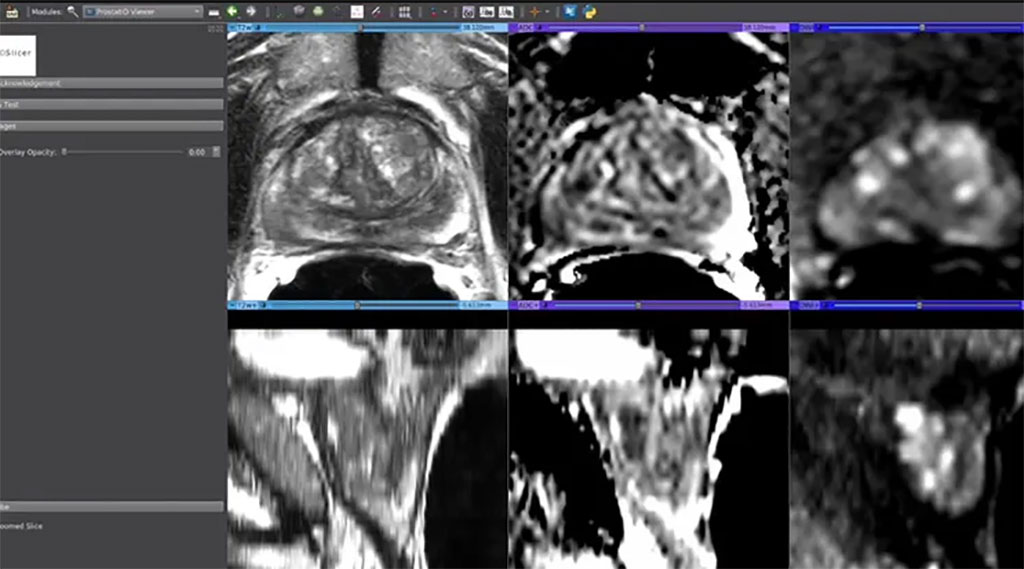
The ProstatID AI software from Bot Image, Inc. (Omaha, NE, USA) is not only intended for cancer detection and diagnosis but also for prostate cancer screening using MRI and a non-invasive/non-contrast "short-form" of MRI sequences known as bi-parametric MRI (bpMRI). bpMRI eliminates the need for expensive, time-consuming contrast agents that can damage the kidneys, thereby facilitating a low cost and easily accessible MRI. Bot Image's Prostate AI has enormous implications for men's health and healthcare cost savings, as it is a highly accurate tool for active surveillance. This means that periodic low-cost MRI scans supplemented with the AI interpretation of ProstatID can be conducted, resulting in significant savings.
In clinical trials conducted by Bot Image, the algorithm performed with 93.6% accuracy when used on its own (with area under the sensitivity-specificity curve). Additionally, physician performance was considerably enhanced. In comparison, no other screening method comes close to matching ProstatID's level of sensitivity, specificity, and ability to locate and determine the extent of cancer. ProstatID has been developed using thousands of proprietary MR image sets, radiologist interpretations, segmented prostate gland image sets, pathology laboratory results, correlation data, and initial AI interpretations to aid in the early diagnosis of prostate cancer. Bot Image has received FDA-clearance for its ProstatID AI software for cancer detection and diagnosis, as well as for prostate cancer screening using MRI and bpMRI.
Related Links:
Bot Image, Inc.
Latest MRI News
- World's First Whole-Body Ultra-High Field MRI Officially Comes To Market
- World's First Sensor Detects Errors in MRI Scans Using Laser Light and Gas
- Diamond Dust Could Offer New Contrast Agent Option for Future MRI Scans
- Combining MRI with PSA Testing Improves Clinical Outcomes for Prostate Cancer Patients
- PET/MRI Improves Diagnostic Accuracy for Prostate Cancer Patients
- Next Generation MR-Guided Focused Ultrasound Ushers In Future of Incisionless Neurosurgery
- Two-Part MRI Scan Detects Prostate Cancer More Quickly without Compromising Diagnostic Quality
- World’s Most Powerful MRI Machine Images Living Brain with Unrivaled Clarity
- New Whole-Body Imaging Technology Makes It Possible to View Inflammation on MRI Scan
- Combining Prostate MRI with Blood Test Can Avoid Unnecessary Prostate Biopsies
- New Treatment Combines MRI and Ultrasound to Control Prostate Cancer without Serious Side Effects
- MRI Improves Diagnosis and Treatment of Prostate Cancer
- Combined PET-MRI Scan Improves Treatment for Early Breast Cancer Patients
- 4D MRI Could Improve Clinical Assessment of Heart Blood Flow Abnormalities
- MRI-Guided Focused Ultrasound Therapy Shows Promise in Treating Prostate Cancer
- AI-Based MRI Tool Outperforms Current Brain Tumor Diagnosis Methods
Channels
Radiography
view channel
Novel Breast Imaging System Proves As Effective As Mammography
Breast cancer remains the most frequently diagnosed cancer among women. It is projected that one in eight women will be diagnosed with breast cancer during her lifetime, and one in 42 women who turn 50... Read more
AI Assistance Improves Breast-Cancer Screening by Reducing False Positives
Radiologists typically detect one case of cancer for every 200 mammograms reviewed. However, these evaluations often result in false positives, leading to unnecessary patient recalls for additional testing,... Read moreUltrasound
view channel
Largest Model Trained On Echocardiography Images Assesses Heart Structure and Function
Foundation models represent an exciting frontier in generative artificial intelligence (AI), yet many lack the specialized medical data needed to make them applicable in healthcare settings.... Read more.jpg)
Groundbreaking Technology Enables Precise, Automatic Measurement of Peripheral Blood Vessels
The current standard of care of using angiographic information is often inadequate for accurately assessing vessel size in the estimated 20 million people in the U.S. who suffer from peripheral vascular disease.... Read more
Deep Learning Advances Super-Resolution Ultrasound Imaging
Ultrasound localization microscopy (ULM) is an advanced imaging technique that offers high-resolution visualization of microvascular structures. It employs microbubbles, FDA-approved contrast agents, injected... Read more
Novel Ultrasound-Launched Targeted Nanoparticle Eliminates Biofilm and Bacterial Infection
Biofilms, formed by bacteria aggregating into dense communities for protection against harsh environmental conditions, are a significant contributor to various infectious diseases. Biofilms frequently... Read moreNuclear Medicine
view channel
New Imaging Technique Monitors Inflammation Disorders without Radiation Exposure
Imaging inflammation using traditional radiological techniques presents significant challenges, including radiation exposure, poor image quality, high costs, and invasive procedures. Now, new contrast... Read more
New SPECT/CT Technique Could Change Imaging Practices and Increase Patient Access
The development of lead-212 (212Pb)-PSMA–based targeted alpha therapy (TAT) is garnering significant interest in treating patients with metastatic castration-resistant prostate cancer. The imaging of 212Pb,... Read moreNew Radiotheranostic System Detects and Treats Ovarian Cancer Noninvasively
Ovarian cancer is the most lethal gynecological cancer, with less than a 30% five-year survival rate for those diagnosed in late stages. Despite surgery and platinum-based chemotherapy being the standard... Read more
AI System Automatically and Reliably Detects Cardiac Amyloidosis Using Scintigraphy Imaging
Cardiac amyloidosis, a condition characterized by the buildup of abnormal protein deposits (amyloids) in the heart muscle, severely affects heart function and can lead to heart failure or death without... Read moreGeneral/Advanced Imaging
view channel
Radiation Therapy Computed Tomography Solution Boosts Imaging Accuracy
One of the most significant challenges in oncology care is disease complexity in terms of the variety of cancer types and the individualized presentation of each patient. This complexity necessitates a... Read more
PET Scans Reveal Hidden Inflammation in Multiple Sclerosis Patients
A key challenge for clinicians treating patients with multiple sclerosis (MS) is that after a certain amount of time, they continue to worsen even though their MRIs show no change. A new study has now... Read moreImaging IT
view channel
New Google Cloud Medical Imaging Suite Makes Imaging Healthcare Data More Accessible
Medical imaging is a critical tool used to diagnose patients, and there are billions of medical images scanned globally each year. Imaging data accounts for about 90% of all healthcare data1 and, until... Read more
Global AI in Medical Diagnostics Market to Be Driven by Demand for Image Recognition in Radiology
The global artificial intelligence (AI) in medical diagnostics market is expanding with early disease detection being one of its key applications and image recognition becoming a compelling consumer proposition... Read moreIndustry News
view channel
Hologic Acquires UK-Based Breast Surgical Guidance Company Endomagnetics Ltd.
Hologic, Inc. (Marlborough, MA, USA) has entered into a definitive agreement to acquire Endomagnetics Ltd. (Cambridge, UK), a privately held developer of breast cancer surgery technologies, for approximately... Read more
Bayer and Google Partner on New AI Product for Radiologists
Medical imaging data comprises around 90% of all healthcare data, and it is a highly complex and rich clinical data modality and serves as a vital tool for diagnosing patients. Each year, billions of medical... Read more


