AI System Quantifies Lung Lesions and Airway Volumes on CT Imaging in IPF Patients
|
By MedImaging International staff writers Posted on 11 Apr 2022 |

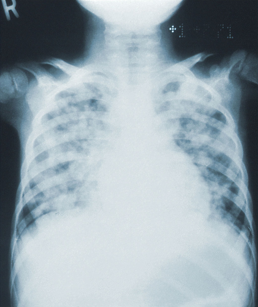
There is a growing need to accurately estimate the prognosis of idiopathic pulmonary fibrosis (IPF) in clinical practice, given the development of effective drugs for treating IPF. Scientists have developed an artificial intelligence (AI)-based image analysis software to detect parenchymal and airway abnormalities on computed tomography (CT) imaging of the chest and to explore their prognostic importance in patients with IPF.
Scientists from the Kyoto University (Kyoto, Japan) developed the novel AI-based quantitative CT image analysis software (AIQCT) by applying 304 high-resolution CT (HRCT) scans from patients with diffuse lung diseases as the training set. AIQCT automatically categorized and quantified 10 types of parenchymal patterns as well as airways, expressing the volumes as percentages of the total lung volume. To validate the software, the area percentages of each lesion quantified by AIQCT were compared with those of the visual scores using 30 plain high-resolution CT images with lung diseases. In addition, three-dimensional analysis for similarity with ground truth was performed using HRCT images from 10 patients with IPF. AIQCT was then applied to 120 patients with IPF who underwent HRCT scanning of the chest at our institute. The associations between the measured volumes and survival were analyzed.
The scientists found that the correlations between AIQCT and the visual scores were moderate to strong (correlation coefficient 0.44–0.95) depending on the parenchymal pattern. The Dice indices for similarity between AIQCT data and ground truth were 0.67, 0.76, and 0.64 for reticulation, honeycombing, and bronchi, respectively. During a median follow-up period of 2,184 days, 66 patients died, and one underwent lung transplantation. In multivariable Cox regression analysis, bronchial volumes (adjusted hazard ratio [HR], 1.33; 95% confidence interval [CI], 1.16–1.53) and normal lung volumes (adjusted HR, 0.97; 95% CI, 0.94–0.99) were independently associated with survival after adjusting for the gender-age-lung physiology stage of IPF.
Based on their findings, the scientists concluded that their newly-developed AI-based image analysis software successfully quantified parenchymal lesions and airway volumes. According to the scientists, bronchial and normal lung volumes on HRCT imaging of the chest can provide additional prognostic information on the gender-age-lung physiology stage of IPF.
Related Links:
Kyoto University
Latest General/Advanced Imaging News
- Radiation Therapy Computed Tomography Solution Boosts Imaging Accuracy
- PET Scans Reveal Hidden Inflammation in Multiple Sclerosis Patients
- Artificial Intelligence Evaluates Cardiovascular Risk from CT Scans
- New AI Method Captures Uncertainty in Medical Images
- CT Coronary Angiography Reduces Need for Invasive Tests to Diagnose Coronary Artery Disease
- Novel Blood Test Could Reduce Need for PET Imaging of Patients with Alzheimer’s
- CT-Based Deep Learning Algorithm Accurately Differentiates Benign From Malignant Vertebral Fractures
- Minimally Invasive Procedure Could Help Patients Avoid Thyroid Surgery
- Self-Driving Mobile C-Arm Reduces Imaging Time during Surgery
- AR Application Turns Medical Scans Into Holograms for Assistance in Surgical Planning
- Imaging Technology Provides Ground-Breaking New Approach for Diagnosing and Treating Bowel Cancer
- CT Coronary Calcium Scoring Predicts Heart Attacks and Strokes
- AI Model Detects 90% of Lymphatic Cancer Cases from PET and CT Images
- Breakthrough Technology Revolutionizes Breast Imaging
- State-Of-The-Art System Enhances Accuracy of Image-Guided Diagnostic and Interventional Procedures
- Catheter-Based Device with New Cardiovascular Imaging Approach Offers Unprecedented View of Dangerous Plaques
Channels
Radiography
view channel
Novel Breast Imaging System Proves As Effective As Mammography
Breast cancer remains the most frequently diagnosed cancer among women. It is projected that one in eight women will be diagnosed with breast cancer during her lifetime, and one in 42 women who turn 50... Read more
AI Assistance Improves Breast-Cancer Screening by Reducing False Positives
Radiologists typically detect one case of cancer for every 200 mammograms reviewed. However, these evaluations often result in false positives, leading to unnecessary patient recalls for additional testing,... Read moreMRI
view channel
Low-Cost Whole-Body MRI Device Combined with AI Generates High-Quality Results
Magnetic Resonance Imaging (MRI) has significantly transformed healthcare, providing a noninvasive, radiation-free method for detailed imaging. It is especially promising for the future of medical diagnosis... Read more
World's First Whole-Body Ultra-High Field MRI Officially Comes To Market
The world's first whole-body ultra-high field (UHF) MRI has officially come to market, marking a remarkable advancement in diagnostic radiology. United Imaging (Shanghai, China) has secured clearance from the U.... Read moreUltrasound
view channel
First AI-Powered POC Ultrasound Diagnostic Solution Helps Prioritize Cases Based On Severity
Ultrasound scans are essential for identifying and diagnosing various medical conditions, but often, patients must wait weeks or months for results due to a shortage of qualified medical professionals... Read more
Largest Model Trained On Echocardiography Images Assesses Heart Structure and Function
Foundation models represent an exciting frontier in generative artificial intelligence (AI), yet many lack the specialized medical data needed to make them applicable in healthcare settings.... Read more.jpg)
Groundbreaking Technology Enables Precise, Automatic Measurement of Peripheral Blood Vessels
The current standard of care of using angiographic information is often inadequate for accurately assessing vessel size in the estimated 20 million people in the U.S. who suffer from peripheral vascular disease.... Read moreNuclear Medicine
view channelNew PET Agent Rapidly and Accurately Visualizes Lesions in Clear Cell Renal Cell Carcinoma Patients
Clear cell renal cell cancer (ccRCC) represents 70-80% of renal cell carcinoma cases. While localized disease can be effectively treated with surgery and ablative therapies, one-third of patients either... Read more
New Imaging Technique Monitors Inflammation Disorders without Radiation Exposure
Imaging inflammation using traditional radiological techniques presents significant challenges, including radiation exposure, poor image quality, high costs, and invasive procedures. Now, new contrast... Read more
New SPECT/CT Technique Could Change Imaging Practices and Increase Patient Access
The development of lead-212 (212Pb)-PSMA–based targeted alpha therapy (TAT) is garnering significant interest in treating patients with metastatic castration-resistant prostate cancer. The imaging of 212Pb,... Read moreImaging IT
view channel
New Google Cloud Medical Imaging Suite Makes Imaging Healthcare Data More Accessible
Medical imaging is a critical tool used to diagnose patients, and there are billions of medical images scanned globally each year. Imaging data accounts for about 90% of all healthcare data1 and, until... Read more
Global AI in Medical Diagnostics Market to Be Driven by Demand for Image Recognition in Radiology
The global artificial intelligence (AI) in medical diagnostics market is expanding with early disease detection being one of its key applications and image recognition becoming a compelling consumer proposition... Read moreIndustry News
view channel
Hologic Acquires UK-Based Breast Surgical Guidance Company Endomagnetics Ltd.
Hologic, Inc. (Marlborough, MA, USA) has entered into a definitive agreement to acquire Endomagnetics Ltd. (Cambridge, UK), a privately held developer of breast cancer surgery technologies, for approximately... Read more
Bayer and Google Partner on New AI Product for Radiologists
Medical imaging data comprises around 90% of all healthcare data, and it is a highly complex and rich clinical data modality and serves as a vital tool for diagnosing patients. Each year, billions of medical... Read more


