Targeted PET Imaging Locates Prostate Cancer and Metastases
|
By MedImaging International staff writers Posted on 28 Sep 2021 |

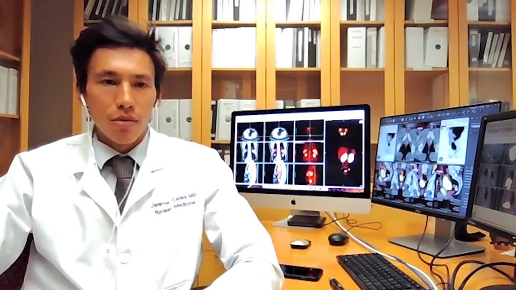
Image: Dr. Jeremie Calais at the 68Ga-PSMA-11 PSMA PET workstation (Photo courtesy of UCLA)
A new positron emission tomography (PET) method can detect prostate-specific membrane antigen (PSMA) radioactive tracers throughout the body, claims a new study.
Developed by researchers at the University of California, San Francisco (UCSF; USA), Aalborg University Hospital (Denmark), the University of California, Los Angeles (UCLA; USA), and other institutions, 68Ga-PSMA-11 is a PSMA imaging tracer for the detection of prostate cancer (PC) nodal metastases. To examine its diagnostic efficacy (as compared with histopathology), they enrolled 764 patients with intermediate- to high-risk PC, 277 of which subsequently underwent radical prostatectomy treatment.
The results revealed that 68Ga-PSMA-11 PET scans were positive in 14% of pelvic nodal cases, one percent of extrapelvic nodal cases, and 3% of patients with bone metastatic disease. Sensitivity for detection of pelvic lymph node metastasis was 40%, indicating that in the remaining 60% of patients, the lesions were too small to be detected (micrometastasis); specificity, however, was 95%, much better than current existing methods. The study was published on September 16, 2021, in JAMA Oncology.
“When a patient is diagnosed with prostate cancer that has some pathologic features on the biopsy that indicate some risk of metastasis in the lymph node or the bones, the physician need to know if the cancer has spread out of the prostate or not,” said senior author Jeremie Calais, MD, of the UCLA department of molecular and medical pharmacology. “PSMA PET/CT is a whole- whole-body imaging modality that can perform a one-time whole body staging with high accuracy for locating and detecting if any metastasis has spread out from the prostate.”
PET is a nuclear medicine imaging technique that produces a three dimensional (3D) image of functional processes in the body. The system detects pairs of gamma rays emitted indirectly by a positron-emitting radionuclide tracer. Tracer concentrations within the body are then constructed in 3D by computer analysis. In modern PET-CT scanners, 3D imaging is often accomplished with the aid of a CT X-ray scan performed on the patient during the same session, in the same machine.
Related Links:
University of California, San Francisco
Aalborg University Hospital
University of California, Los Angeles
Latest General/Advanced Imaging News
- Bone Density Test Uses Existing CT Images to Predict Fractures
- AI Predicts Cardiac Risk and Mortality from Routine Chest CT Scans
- Radiation Therapy Computed Tomography Solution Boosts Imaging Accuracy
- PET Scans Reveal Hidden Inflammation in Multiple Sclerosis Patients
- Artificial Intelligence Evaluates Cardiovascular Risk from CT Scans
- New AI Method Captures Uncertainty in Medical Images
- CT Coronary Angiography Reduces Need for Invasive Tests to Diagnose Coronary Artery Disease
- Novel Blood Test Could Reduce Need for PET Imaging of Patients with Alzheimer’s
- CT-Based Deep Learning Algorithm Accurately Differentiates Benign From Malignant Vertebral Fractures
- Minimally Invasive Procedure Could Help Patients Avoid Thyroid Surgery
- Self-Driving Mobile C-Arm Reduces Imaging Time during Surgery
- AR Application Turns Medical Scans Into Holograms for Assistance in Surgical Planning
- Imaging Technology Provides Ground-Breaking New Approach for Diagnosing and Treating Bowel Cancer
- CT Coronary Calcium Scoring Predicts Heart Attacks and Strokes
- AI Model Detects 90% of Lymphatic Cancer Cases from PET and CT Images
- Breakthrough Technology Revolutionizes Breast Imaging
Channels
Radiography
view channel
Novel Breast Imaging System Proves As Effective As Mammography
Breast cancer remains the most frequently diagnosed cancer among women. It is projected that one in eight women will be diagnosed with breast cancer during her lifetime, and one in 42 women who turn 50... Read more
AI Assistance Improves Breast-Cancer Screening by Reducing False Positives
Radiologists typically detect one case of cancer for every 200 mammograms reviewed. However, these evaluations often result in false positives, leading to unnecessary patient recalls for additional testing,... Read moreMRI
view channel
Low-Cost Whole-Body MRI Device Combined with AI Generates High-Quality Results
Magnetic Resonance Imaging (MRI) has significantly transformed healthcare, providing a noninvasive, radiation-free method for detailed imaging. It is especially promising for the future of medical diagnosis... Read more
World's First Whole-Body Ultra-High Field MRI Officially Comes To Market
The world's first whole-body ultra-high field (UHF) MRI has officially come to market, marking a remarkable advancement in diagnostic radiology. United Imaging (Shanghai, China) has secured clearance from the U.... Read moreUltrasound
view channel.jpg)
Diagnostic System Automatically Analyzes TTE Images to Identify Congenital Heart Disease
Congenital heart disease (CHD) is one of the most prevalent congenital anomalies worldwide, presenting substantial health and financial challenges for affected patients. Early detection and treatment of... Read more
Super-Resolution Imaging Technique Could Improve Evaluation of Cardiac Conditions
The heart depends on efficient blood circulation to pump blood throughout the body, delivering oxygen to tissues and removing carbon dioxide and waste. Yet, when heart vessels are damaged, it can disrupt... Read more
First AI-Powered POC Ultrasound Diagnostic Solution Helps Prioritize Cases Based On Severity
Ultrasound scans are essential for identifying and diagnosing various medical conditions, but often, patients must wait weeks or months for results due to a shortage of qualified medical professionals... Read moreNuclear Medicine
view channel
New PET Biomarker Predicts Success of Immune Checkpoint Blockade Therapy
Immunotherapies, such as immune checkpoint blockade (ICB), have shown promising clinical results in treating melanoma, non-small cell lung cancer, and other tumor types. However, the effectiveness of these... Read moreNew PET Agent Rapidly and Accurately Visualizes Lesions in Clear Cell Renal Cell Carcinoma Patients
Clear cell renal cell cancer (ccRCC) represents 70-80% of renal cell carcinoma cases. While localized disease can be effectively treated with surgery and ablative therapies, one-third of patients either... Read more
New Imaging Technique Monitors Inflammation Disorders without Radiation Exposure
Imaging inflammation using traditional radiological techniques presents significant challenges, including radiation exposure, poor image quality, high costs, and invasive procedures. Now, new contrast... Read more
New SPECT/CT Technique Could Change Imaging Practices and Increase Patient Access
The development of lead-212 (212Pb)-PSMA–based targeted alpha therapy (TAT) is garnering significant interest in treating patients with metastatic castration-resistant prostate cancer. The imaging of 212Pb,... Read moreImaging IT
view channel
New Google Cloud Medical Imaging Suite Makes Imaging Healthcare Data More Accessible
Medical imaging is a critical tool used to diagnose patients, and there are billions of medical images scanned globally each year. Imaging data accounts for about 90% of all healthcare data1 and, until... Read more
Global AI in Medical Diagnostics Market to Be Driven by Demand for Image Recognition in Radiology
The global artificial intelligence (AI) in medical diagnostics market is expanding with early disease detection being one of its key applications and image recognition becoming a compelling consumer proposition... Read moreIndustry News
view channel
Hologic Acquires UK-Based Breast Surgical Guidance Company Endomagnetics Ltd.
Hologic, Inc. (Marlborough, MA, USA) has entered into a definitive agreement to acquire Endomagnetics Ltd. (Cambridge, UK), a privately held developer of breast cancer surgery technologies, for approximately... Read more
Bayer and Google Partner on New AI Product for Radiologists
Medical imaging data comprises around 90% of all healthcare data, and it is a highly complex and rich clinical data modality and serves as a vital tool for diagnosing patients. Each year, billions of medical... Read more


