PET Agent May Gauge Breast Cancer Therapy Efficacy
|
By MedImaging International staff writers Posted on 27 Feb 2019 |

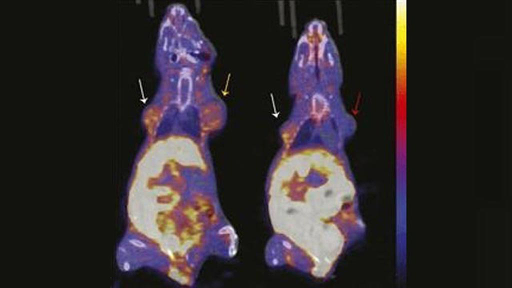
Image: Representative images of coronal 18F-FFNP uptake on PET/CT (Photo courtesy of UWHealth).
Physicians may soon have a new way to measure the efficacy or failure of hormone therapy for breast cancer patients, according to a new study.
Researchers at the University of Wisconsin School of Medicine and Public Health (UWHealth; Madison, USA) conducted a study to evaluate the ability of positron emission tomography (PET) imaging with 18F-fluorofuranylnorprogesterone (18F-FFNP) to measure changes in progesterone receptor (PR) levels resulting from an estradiol challenge, which can determine the likelihood of potential benefit of hormonal therapies targeting estrogen receptor for individual patients.
For the study, cultured T47D human breast cancer cells and mice bearing T47D tumor xenografts were treated with estrogen to increase PR expression. The cells and mice were then imaged with 18F-FFNP, and assays were conducted for cell uptake and tissue biodistribution. In vitro 18F-FFNP binding was measured by saturation and competitive binding assays, while in vivo uptake was measured with PET imaging.
The results revealed that in T47D cells treated with estrogen, an increase in 18F-FFNP uptake was measured at 48 hours after treatment; in mice with T47D tumor xenografts, increased uptake was seen at 48 and 72 hours after treatment. The increase in 18F-FFNP uptake also correlated with an increase in PR protein expression and proliferation. The study was published in the February 2019 issue of The Journal of Nuclear Medicine.
“Validation of PR imaging as a biomarker of endocrine sensitivity in patients before and after estradiol challenge could provide new opportunities in the field of molecular imaging and nuclear medicine for breast cancer imaging,” said senior author Amy Fowler, MD, PhD, of the UWHealth department of radiology. “Improved methods for testing endocrine sensitivity in patients could better inform decisions for optimal individualized ER-positive breast cancer therapy, potentially reducing morbidity and mortality.”
ER-positive breast cancer is the most common class of breast cancer, affecting nearly 70% of patients. By participating in an estradiol challenge, physicians can determine the likelihood of potential benefit of hormonal therapies targeting ER for individual patients. Many hormone therapies interfere with the ability of estrogen to regulate the expression of PR protein, which is more pronounced in the presence of estrogen. As such, several PET tracers have been developed to monitor and analyze changes in the PR level during therapy.
Related Links:
University of Wisconsin School of Medicine and Public Health
Latest General/Advanced Imaging News
- Bone Density Test Uses Existing CT Images to Predict Fractures
- AI Predicts Cardiac Risk and Mortality from Routine Chest CT Scans
- Radiation Therapy Computed Tomography Solution Boosts Imaging Accuracy
- PET Scans Reveal Hidden Inflammation in Multiple Sclerosis Patients
- Artificial Intelligence Evaluates Cardiovascular Risk from CT Scans
- New AI Method Captures Uncertainty in Medical Images
- CT Coronary Angiography Reduces Need for Invasive Tests to Diagnose Coronary Artery Disease
- Novel Blood Test Could Reduce Need for PET Imaging of Patients with Alzheimer’s
- CT-Based Deep Learning Algorithm Accurately Differentiates Benign From Malignant Vertebral Fractures
- Minimally Invasive Procedure Could Help Patients Avoid Thyroid Surgery
- Self-Driving Mobile C-Arm Reduces Imaging Time during Surgery
- AR Application Turns Medical Scans Into Holograms for Assistance in Surgical Planning
- Imaging Technology Provides Ground-Breaking New Approach for Diagnosing and Treating Bowel Cancer
- CT Coronary Calcium Scoring Predicts Heart Attacks and Strokes
- AI Model Detects 90% of Lymphatic Cancer Cases from PET and CT Images
- Breakthrough Technology Revolutionizes Breast Imaging
Channels
Radiography
view channel
Novel Breast Imaging System Proves As Effective As Mammography
Breast cancer remains the most frequently diagnosed cancer among women. It is projected that one in eight women will be diagnosed with breast cancer during her lifetime, and one in 42 women who turn 50... Read more
AI Assistance Improves Breast-Cancer Screening by Reducing False Positives
Radiologists typically detect one case of cancer for every 200 mammograms reviewed. However, these evaluations often result in false positives, leading to unnecessary patient recalls for additional testing,... Read moreMRI
view channel
Low-Cost Whole-Body MRI Device Combined with AI Generates High-Quality Results
Magnetic Resonance Imaging (MRI) has significantly transformed healthcare, providing a noninvasive, radiation-free method for detailed imaging. It is especially promising for the future of medical diagnosis... Read more
World's First Whole-Body Ultra-High Field MRI Officially Comes To Market
The world's first whole-body ultra-high field (UHF) MRI has officially come to market, marking a remarkable advancement in diagnostic radiology. United Imaging (Shanghai, China) has secured clearance from the U.... Read moreUltrasound
view channel.jpg)
Diagnostic System Automatically Analyzes TTE Images to Identify Congenital Heart Disease
Congenital heart disease (CHD) is one of the most prevalent congenital anomalies worldwide, presenting substantial health and financial challenges for affected patients. Early detection and treatment of... Read more
Super-Resolution Imaging Technique Could Improve Evaluation of Cardiac Conditions
The heart depends on efficient blood circulation to pump blood throughout the body, delivering oxygen to tissues and removing carbon dioxide and waste. Yet, when heart vessels are damaged, it can disrupt... Read more
First AI-Powered POC Ultrasound Diagnostic Solution Helps Prioritize Cases Based On Severity
Ultrasound scans are essential for identifying and diagnosing various medical conditions, but often, patients must wait weeks or months for results due to a shortage of qualified medical professionals... Read moreNuclear Medicine
view channel
New PET Biomarker Predicts Success of Immune Checkpoint Blockade Therapy
Immunotherapies, such as immune checkpoint blockade (ICB), have shown promising clinical results in treating melanoma, non-small cell lung cancer, and other tumor types. However, the effectiveness of these... Read moreNew PET Agent Rapidly and Accurately Visualizes Lesions in Clear Cell Renal Cell Carcinoma Patients
Clear cell renal cell cancer (ccRCC) represents 70-80% of renal cell carcinoma cases. While localized disease can be effectively treated with surgery and ablative therapies, one-third of patients either... Read more
New Imaging Technique Monitors Inflammation Disorders without Radiation Exposure
Imaging inflammation using traditional radiological techniques presents significant challenges, including radiation exposure, poor image quality, high costs, and invasive procedures. Now, new contrast... Read more
New SPECT/CT Technique Could Change Imaging Practices and Increase Patient Access
The development of lead-212 (212Pb)-PSMA–based targeted alpha therapy (TAT) is garnering significant interest in treating patients with metastatic castration-resistant prostate cancer. The imaging of 212Pb,... Read moreImaging IT
view channel
New Google Cloud Medical Imaging Suite Makes Imaging Healthcare Data More Accessible
Medical imaging is a critical tool used to diagnose patients, and there are billions of medical images scanned globally each year. Imaging data accounts for about 90% of all healthcare data1 and, until... Read more
Global AI in Medical Diagnostics Market to Be Driven by Demand for Image Recognition in Radiology
The global artificial intelligence (AI) in medical diagnostics market is expanding with early disease detection being one of its key applications and image recognition becoming a compelling consumer proposition... Read moreIndustry News
view channel
Hologic Acquires UK-Based Breast Surgical Guidance Company Endomagnetics Ltd.
Hologic, Inc. (Marlborough, MA, USA) has entered into a definitive agreement to acquire Endomagnetics Ltd. (Cambridge, UK), a privately held developer of breast cancer surgery technologies, for approximately... Read more
Bayer and Google Partner on New AI Product for Radiologists
Medical imaging data comprises around 90% of all healthcare data, and it is a highly complex and rich clinical data modality and serves as a vital tool for diagnosing patients. Each year, billions of medical... Read more


