Sugar Could Replace Contrast Agents in Brain MRI Scans
|
By MedImaging International staff writers Posted on 05 Jul 2017 |

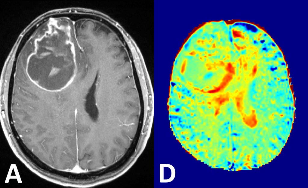
Image: Visualization of brain cancer by conventional contrast agents (L) and using glucose MRI method (R) (Photo courtesy of DKFZ).
A new study suggests that glucose metabolism activity identified using magnetic resonance imaging (MRI) could help identify solid tumors or very aggressively growing tumor areas.
Researchers at the German Cancer Research Center (DKFZ; Heidelberg, Germany), the Max-Planck-Institute for Biological Cybernetics (Tuebingen, Germany), and other institutions used an ultrahigh field 7 Tesla MRI scanner to detect intracerebral regions of increased glucose concentration. For the study, they examined nine patients with newly diagnosed glioblastoma and four healthy volunteers between October 2015 and July 2016.
T1ρ-weighted dynamic glucose-enhanced (DGE) MRI was performed with injection of 100 mL of 20% d-glucose via the cubital vein. Glucose enhancement, given by the relative signal intensity change, was then quantitatively investigated in brain gray matter versus white matter in the healthy volunteers, and in tumor tissue versus normal-appearing white matter of patients with glioblastoma. The median signal intensities of the assessed brain regions were then compared.
The results demonstrated that in healthy volunteers, the median signal intensity in basal ganglia gray matter was significantly increased compared with that in white matter tissue. But in the glioblastoma patients, the median signal intensity in the glucose-enhanced tumor region, as displayed on the T1ρ-weighted DGE images, was significantly higher than that in contralateral normal-appearing white matter, indicating the pathophysiologically increased glucose uptake that occurs in human brain cancer. The study was published on June 23, 2017, in Radiology.
“We do not know yet how the shares of measured glucose are distributed between vessels and extracellular spaces on the one hand, and the cell interior on the other,” said study co-author radiologist Heinz-Peter Schlemmer, MD, PhD, of the DKFZ division of radiology. “If we can confirm that substantial signal levels originate from glucose in the cell interior, this would be important additional information for tumor imaging and functional MRI. This could enhance therapy planning and monitoring.”
MRI contrast agents enhance signals in blood vessels and in spaces between cells, but do not reach the interior of the cell; by contrast, glucose is taken up and then broken down in the body cells, and tumor cells are particularly hungry for glucose in order to feed their high-energy needs. As glucose is found at much lower levels in the body, magnetization transfer effect is used to reinforce the glucose signal by transferring the signal from glucose protons to bodily water, which is measured in MRI. The amount of glucose needed corresponds to about five sugar cubes.
Related Links:
German Cancer Research Center
Max-Planck-Institute for Biological Cybernetics
Latest MRI News
- Diamond Dust Could Offer New Contrast Agent Option for Future MRI Scans
- Combining MRI with PSA Testing Improves Clinical Outcomes for Prostate Cancer Patients
- PET/MRI Improves Diagnostic Accuracy for Prostate Cancer Patients
- Next Generation MR-Guided Focused Ultrasound Ushers In Future of Incisionless Neurosurgery
- Two-Part MRI Scan Detects Prostate Cancer More Quickly without Compromising Diagnostic Quality
- World’s Most Powerful MRI Machine Images Living Brain with Unrivaled Clarity
- New Whole-Body Imaging Technology Makes It Possible to View Inflammation on MRI Scan
- Combining Prostate MRI with Blood Test Can Avoid Unnecessary Prostate Biopsies
- New Treatment Combines MRI and Ultrasound to Control Prostate Cancer without Serious Side Effects
- MRI Improves Diagnosis and Treatment of Prostate Cancer
- Combined PET-MRI Scan Improves Treatment for Early Breast Cancer Patients
- 4D MRI Could Improve Clinical Assessment of Heart Blood Flow Abnormalities
- MRI-Guided Focused Ultrasound Therapy Shows Promise in Treating Prostate Cancer
- AI-Based MRI Tool Outperforms Current Brain Tumor Diagnosis Methods
- DW-MRI Lights up Small Ovarian Lesions like Light Bulbs
- Abbreviated Breast MRI Effective for High-Risk Screening without Compromising Diagnostic Accuracy
Channels
Radiography
view channel
Novel Breast Imaging System Proves As Effective As Mammography
Breast cancer remains the most frequently diagnosed cancer among women. It is projected that one in eight women will be diagnosed with breast cancer during her lifetime, and one in 42 women who turn 50... Read more
AI Assistance Improves Breast-Cancer Screening by Reducing False Positives
Radiologists typically detect one case of cancer for every 200 mammograms reviewed. However, these evaluations often result in false positives, leading to unnecessary patient recalls for additional testing,... Read moreUltrasound
view channel.jpg)
Groundbreaking Technology Enables Precise, Automatic Measurement of Peripheral Blood Vessels
The current standard of care of using angiographic information is often inadequate for accurately assessing vessel size in the estimated 20 million people in the U.S. who suffer from peripheral vascular disease.... Read more
Deep Learning Advances Super-Resolution Ultrasound Imaging
Ultrasound localization microscopy (ULM) is an advanced imaging technique that offers high-resolution visualization of microvascular structures. It employs microbubbles, FDA-approved contrast agents, injected... Read more
Novel Ultrasound-Launched Targeted Nanoparticle Eliminates Biofilm and Bacterial Infection
Biofilms, formed by bacteria aggregating into dense communities for protection against harsh environmental conditions, are a significant contributor to various infectious diseases. Biofilms frequently... Read moreNuclear Medicine
view channel
New Imaging Technique Monitors Inflammation Disorders without Radiation Exposure
Imaging inflammation using traditional radiological techniques presents significant challenges, including radiation exposure, poor image quality, high costs, and invasive procedures. Now, new contrast... Read more
New SPECT/CT Technique Could Change Imaging Practices and Increase Patient Access
The development of lead-212 (212Pb)-PSMA–based targeted alpha therapy (TAT) is garnering significant interest in treating patients with metastatic castration-resistant prostate cancer. The imaging of 212Pb,... Read moreNew Radiotheranostic System Detects and Treats Ovarian Cancer Noninvasively
Ovarian cancer is the most lethal gynecological cancer, with less than a 30% five-year survival rate for those diagnosed in late stages. Despite surgery and platinum-based chemotherapy being the standard... Read more
AI System Automatically and Reliably Detects Cardiac Amyloidosis Using Scintigraphy Imaging
Cardiac amyloidosis, a condition characterized by the buildup of abnormal protein deposits (amyloids) in the heart muscle, severely affects heart function and can lead to heart failure or death without... Read moreGeneral/Advanced Imaging
view channel
PET Scans Reveal Hidden Inflammation in Multiple Sclerosis Patients
A key challenge for clinicians treating patients with multiple sclerosis (MS) is that after a certain amount of time, they continue to worsen even though their MRIs show no change. A new study has now... Read more
Artificial Intelligence Evaluates Cardiovascular Risk from CT Scans
Chest computed tomography (CT) is a common diagnostic tool, with approximately 15 million scans conducted each year in the United States, though many are underutilized or not fully explored.... Read more
New AI Method Captures Uncertainty in Medical Images
In the field of biomedicine, segmentation is the process of annotating pixels from an important structure in medical images, such as organs or cells. Artificial Intelligence (AI) models are utilized to... Read more.jpg)
CT Coronary Angiography Reduces Need for Invasive Tests to Diagnose Coronary Artery Disease
Coronary artery disease (CAD), one of the leading causes of death worldwide, involves the narrowing of coronary arteries due to atherosclerosis, resulting in insufficient blood flow to the heart muscle.... Read moreImaging IT
view channel
New Google Cloud Medical Imaging Suite Makes Imaging Healthcare Data More Accessible
Medical imaging is a critical tool used to diagnose patients, and there are billions of medical images scanned globally each year. Imaging data accounts for about 90% of all healthcare data1 and, until... Read more
Global AI in Medical Diagnostics Market to Be Driven by Demand for Image Recognition in Radiology
The global artificial intelligence (AI) in medical diagnostics market is expanding with early disease detection being one of its key applications and image recognition becoming a compelling consumer proposition... Read moreIndustry News
view channel
Bayer and Google Partner on New AI Product for Radiologists
Medical imaging data comprises around 90% of all healthcare data, and it is a highly complex and rich clinical data modality and serves as a vital tool for diagnosing patients. Each year, billions of medical... Read more


